 Movie
Movie Controller

[English] 日本語
 Yorodumi
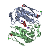
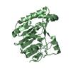
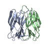
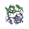
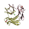
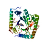
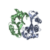
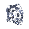
- PDB-1g0z: SPECIFIC MUTATIONS IN KRAIT PLA2 LEAD TO DIMERIZATION OF PROTEIN ... -
+ Open data

- Basic information
| Entry | Database: PDB / ID: 1g0z | ||||||
|---|---|---|---|---|---|---|---|
| Title | SPECIFIC MUTATIONS IN KRAIT PLA2 LEAD TO DIMERIZATION OF PROTEIN MOLECULES: CRYSTAL STRUCTURE OF KRAIT PLA2 AT 2.1 RESOLUTION | ||||||
 Components Components | PHOSPHOLIPASE A2 | ||||||
 Keywords Keywords | TOXIN / Phospholipase A2 / Homodimer / Bungarus caeruleus | ||||||
| Function / homology |  Function and homology informationphospholipase A2 / phospholipase A2 activity / arachidonic acid secretion / phospholipid metabolic process / lipid catabolic process / toxin activity / calcium ion binding / extracellular region Function and homology informationphospholipase A2 / phospholipase A2 activity / arachidonic acid secretion / phospholipid metabolic process / lipid catabolic process / toxin activity / calcium ion binding / extracellular regionSimilarity search - Function | ||||||
| Biological species |  Bungarus caeruleus (cobra) Bungarus caeruleus (cobra) | ||||||
| Method | X-RAY DIFFRACTION / SYNCHROTRON / MOLECULAR REPLACEMENT / Resolution: 2.18 Å | ||||||
 Authors Authors | Singh, T.P. / Gourinath, S. / Sharma, S. / Singh, G. | ||||||
 Citation Citation | Journal: To be Published Title: Specific mutations in Krait PLA2 lead to dimerization of protein molecules: Crystal structure of Krait PLA2 at 2.1 resolution. Authors: Gourinath, S. / Singh, G. / Sharma, S. / Bhanumathi, S. / Betzel, C. / Paramsivam, M. / Srinivasan, A. / Singh, T.P. #1: Journal: To be PublishedTitle: Three-dimensional structure of a novel phospholipase A2 from Indian common krait at 2.45 resolution. Authors: Singh, G. / Gourinath, S. / Sharma, S. / Paramasivam, M. / Srinivasan, A. / Singh, T.P. | ||||||
| History |
|
- Structure visualization
| Structure viewer | Molecule: MolmilJmol/JSmol |
|---|
- Downloads & links
-Download
| PDBx/mmCIF format | 1g0z.cif.gz | 61.5 KB | Display | PDBx/mmCIF format |
|---|---|---|---|---|
| PDB format | pdb1g0z.ent.gz | 44.9 KB | Display | PDB format |
| PDBx/mmJSON format | 1g0z.json.gz | Tree view | PDBx/mmJSON format | |
| Others |  Other downloads Other downloads |
-Validation report
| Arichive directory | https://data.pdbj.org/pub/pdb/validation_reports/g0/1g0zftp://data.pdbj.org/pub/pdb/validation_reports/g0/1g0z | HTTPS FTP |
|---|
-Related structure data
| Related structure data | 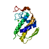 1fe5S S: Starting model for refinement |
|---|---|
| Similar structure data |


-Links
 PDBj
PDBj

- Assembly
| Deposited unit | 
| ||||||||
|---|---|---|---|---|---|---|---|---|---|
| 1 |
| ||||||||
| Unit cell |
|
-Components
| #1: Protein | Mass: 12980.627 Da / Num. of mol.: 2 / Fragment: RESIDUES 28-145 / Source method: isolated from a natural source / Details: NATURAL PROTEIN-ISOFORM / Source: (natural) Bungarus caeruleus (cobra) / Secretion: VENOM / References: UniProt: Q6SLM1*PLUS, phospholipase A2#2: Chemical | Chloride  Mass: 35.453 Da / Num. of mol.: 2 / Source method: obtained synthetically / Formula: Cl Mass: 35.453 Da / Num. of mol.: 2 / Source method: obtained synthetically / Formula: Cl#3: Water | ChemComp-HOH / | Water Mass: 18.015 Da / Num. of mol.: 213 / Source method: isolated from a natural source / Formula: H2O Mass: 18.015 Da / Num. of mol.: 213 / Source method: isolated from a natural source / Formula: H2O |
|---|
-Experimental details
-Experiment
| Experiment | Method: X-RAY DIFFRACTION / Number of used crystals: 1 |
|---|
- Sample preparation
| Crystal | Density Matthews: 2.4 Å3/Da / Density % sol: 48 % |
|---|---|
| Crystal grow | Temperature: 281 K / Method: vapor diffusion, sitting drop / pH: 8.5 Details: 50 mM Tris. HCl, 2.8 M NaCl, 1mM NaN3., pH 8.5, VAPOR DIFFUSION, SITTING DROP, temperature 281K |
-Data collection
| Diffraction | Mean temperature: 180 K |
|---|---|
| Diffraction source | Source: SYNCHROTRON / Site: EMBL/DESY, HAMBURG  / Beamline: X11 / Wavelength: 0.98 / Beamline: X11 / Wavelength: 0.98 |
| Detector | Type: MARRESEARCH / Detector: IMAGE PLATE / Date: Aug 22, 2000 |
| Radiation | Protocol: SINGLE WAVELENGTH / Monochromatic (M) / Laue (L): M / Scattering type: x-ray |
| Radiation wavelength | Wavelength: 0.98 Å / Relative weight: 1 |
| Reflection | Resolution: 2.1→14.3 Å / Num. all: 12541 / Num. obs: 12475 / % possible obs: 99.6 % / Observed criterion σ(F): 0 / Observed criterion σ(I): -3 / Redundancy: 7.8 % / Biso Wilson estimate: 24.7 Å2 / Rsym value: 0.126 / Net I/σ(I): 6.4 |
| Reflection shell | Resolution: 2.18→2.26 Å / Redundancy: 7.8 % / Rsym value: 0.24 / % possible all: 99.2 |
- Processing
| Software |
| ||||||||||||||||||||||||||||||||||||||||||||||||||||||||||||
|---|---|---|---|---|---|---|---|---|---|---|---|---|---|---|---|---|---|---|---|---|---|---|---|---|---|---|---|---|---|---|---|---|---|---|---|---|---|---|---|---|---|---|---|---|---|---|---|---|---|---|---|---|---|---|---|---|---|---|---|---|---|
| Refinement | Method to determine structure: MOLECULAR REPLACEMENT Starting model: PDB ID 1FE5 Resolution: 2.18→14.26 Å / Rfactor Rfree error: 0.01 / Data cutoff high absF: 48393.95 / Data cutoff low absF: 0 / Isotropic thermal model: RESTRAINED / Cross valid method: THROUGHOUT / σ(F): 0 / σ(I): 0 / Stereochemistry target values: Engh & Huber / Details: Used weighted full matrix least squares procedure.
| ||||||||||||||||||||||||||||||||||||||||||||||||||||||||||||
| Solvent computation | Solvent model: FLAT MODEL / Bsol: 80.2238 Å2 / ksol: 0.43391 e/Å3 | ||||||||||||||||||||||||||||||||||||||||||||||||||||||||||||
| Displacement parameters | Biso mean: 24.8 Å2
| ||||||||||||||||||||||||||||||||||||||||||||||||||||||||||||
| Refine analyze |
| ||||||||||||||||||||||||||||||||||||||||||||||||||||||||||||
| Refinement step | Cycle: LAST / Resolution: 2.18→14.26 Å
| ||||||||||||||||||||||||||||||||||||||||||||||||||||||||||||
| Refine LS restraints |
| ||||||||||||||||||||||||||||||||||||||||||||||||||||||||||||
| LS refinement shell | Resolution: 2.18→2.32 Å / Rfactor Rfree error: 0.032 / Total num. of bins used: 6
| ||||||||||||||||||||||||||||||||||||||||||||||||||||||||||||
| Xplor file |
|
