 Movie
Movie Controller

[English] 日本語
 Yorodumi
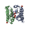
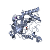
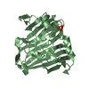
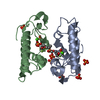
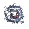
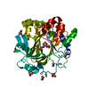
- PDB-1fx9: CARBOXYLIC ESTER HYDROLASE COMPLEX (DIMERIC PLA2 + MJ33 INHIBITOR... -
+ Open data

- Basic information
| Entry | Database: PDB / ID: 1fx9 | ||||||
|---|---|---|---|---|---|---|---|
| Title | CARBOXYLIC ESTER HYDROLASE COMPLEX (DIMERIC PLA2 + MJ33 INHIBITOR + SULPHATE IONS) | ||||||
 Components Components | PHOSPHOLIPASE A2, MAJOR ISOENZYME | ||||||
 Keywords Keywords | HYDROLASE / ENZYME / CARBOXYLIC ESTER HYDROLASE / DIMER / SULPHATE BINDING / INHIBITOR BINDING | ||||||
| Function / homology |  Function and homology information Function and homology informationAcyl chain remodelling of PC / Acyl chain remodelling of PS / Acyl chain remodelling of PE / Acyl chain remodelling of PI / Acyl chain remodelling of PG / Synthesis of PA / positive regulation of podocyte apoptotic process / regulation of glucose import / phosphatidylglycerol metabolic process / calcium-dependent phospholipase A2 activity ...Acyl chain remodelling of PC / Acyl chain remodelling of PS / Acyl chain remodelling of PE / Acyl chain remodelling of PI / Acyl chain remodelling of PG / Synthesis of PA / positive regulation of podocyte apoptotic process / regulation of glucose import / phosphatidylglycerol metabolic process / calcium-dependent phospholipase A2 activity / phosphatidylcholine metabolic process / leukotriene biosynthetic process / neutrophil mediated immunity / phospholipase A2 / bile acid binding / phospholipase A2 activity / positive regulation of calcium ion transport into cytosol / phospholipid metabolic process / lipid catabolic process / neutrophil chemotaxis / positive regulation of interleukin-8 production / phospholipid binding / positive regulation of MAP kinase activity / cellular response to insulin stimulus / fatty acid biosynthetic process / positive regulation of fibroblast proliferation / positive regulation of immune response / positive regulation of NF-kappaB transcription factor activity / intracellular signal transduction / signaling receptor binding / calcium ion binding / positive regulation of cell population proliferation / cell surface / positive regulation of transcription by RNA polymerase II / extracellular regionSimilarity search - Function | ||||||
| Biological species |  Sus scrofa (pig) Sus scrofa (pig) | ||||||
| Method | X-RAY DIFFRACTION / MOLECULAR REPLACEMENT / Resolution: 2 Å | ||||||
 Authors Authors | Pan, Y.H. / Epstein, T.M. / Jain, M.K. / Bahnson, B.J. | ||||||
 Citation Citation | Journal: Biochemistry / Year: 2001 Title: Five coplanar anion binding sites on one face of phospholipase A2: relationship to interface binding. Authors: Pan, Y.H. / Epstein, T.M. / Jain, M.K. / Bahnson, B.J. | ||||||
| History |
|
- Structure visualization
| Structure viewer | Molecule: MolmilJmol/JSmol |
|---|
- Downloads & links
-Download
| PDBx/mmCIF format | 1fx9.cif.gz | 66.8 KB | Display | PDBx/mmCIF format |
|---|---|---|---|---|
| PDB format | pdb1fx9.ent.gz | 49.4 KB | Display | PDB format |
| PDBx/mmJSON format | 1fx9.json.gz | Tree view | PDBx/mmJSON format | |
| Others |  Other downloads Other downloads |
-Validation report
| Arichive directory | https://data.pdbj.org/pub/pdb/validation_reports/fx/1fx9ftp://data.pdbj.org/pub/pdb/validation_reports/fx/1fx9 | HTTPS FTP |
|---|
-Related structure data
| Related structure data | 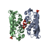 1fxfC  4p2pS C: citing same article ( S: Starting model for refinement |
|---|---|
| Similar structure data |

-Links
 PDBj
PDBj

- Assembly
| Deposited unit | 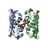
| ||||||||
|---|---|---|---|---|---|---|---|---|---|
| 1 |
| ||||||||
| Unit cell |
|
-Components
| #1: Protein | / PHOSPHATIDYLCHOLINE 2-ACYLHYDROLASE / PLA2 Mass: 14009.714 Da / Num. of mol.: 2 Source method: isolated from a genetically manipulated source Source: (gene. exp.) Sus scrofa (pig) / Production host:  Escherichia coli (E. coli) / References: UniProt: P00592, phospholipase A2 Escherichia coli (E. coli) / References: UniProt: P00592, phospholipase A2#2: Chemical |   Mass: 40.078 Da / Num. of mol.: 2 / Source method: obtained synthetically / Formula: Ca Mass: 40.078 Da / Num. of mol.: 2 / Source method: obtained synthetically / Formula: Ca#3: Chemical | ChemComp-SO4 / Sulfate  Mass: 96.063 Da / Num. of mol.: 5 / Source method: obtained synthetically / Formula: SO4 Mass: 96.063 Da / Num. of mol.: 5 / Source method: obtained synthetically / Formula: SO4#4: Chemical | ChemComp-MJI / | 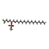  Mass: 492.550 Da / Num. of mol.: 1 / Source method: obtained synthetically / Formula: C22H44F3O6P Mass: 492.550 Da / Num. of mol.: 1 / Source method: obtained synthetically / Formula: C22H44F3O6P#5: Water | ChemComp-HOH / | Water Mass: 18.015 Da / Num. of mol.: 185 / Source method: isolated from a natural source / Formula: H2O Mass: 18.015 Da / Num. of mol.: 185 / Source method: isolated from a natural source / Formula: H2O |
|---|
-Experimental details
-Experiment
| Experiment | Method: X-RAY DIFFRACTION / Number of used crystals: 1 |
|---|
- Sample preparation
| Crystal | Density Matthews: 2 Å3/Da / Density % sol: 54.95 % | ||||||||||||||||||||||||||||||||||||||||||
|---|---|---|---|---|---|---|---|---|---|---|---|---|---|---|---|---|---|---|---|---|---|---|---|---|---|---|---|---|---|---|---|---|---|---|---|---|---|---|---|---|---|---|---|
| Crystal grow | Temperature: 298 K / Method: evaporation / pH: 4.6 Details: 25% PEG3500, 0.2 M SODIUM SULPHATE, 0.1 M SODIUM ACETATE BUFFER, PH 4.6, EVAPORATION, temperature 298K | ||||||||||||||||||||||||||||||||||||||||||
| Crystal grow | *PLUS Temperature: 25 ℃ / Method: vapor diffusion, hanging drop | ||||||||||||||||||||||||||||||||||||||||||
| Components of the solutions | *PLUS
|
-Data collection
| Diffraction | Mean temperature: 93 K |
|---|---|
| Diffraction source | Source: ROTATING ANODE / Type: RIGAKU RUH3R / Wavelength: 1.5418 |
| Detector | Type: RIGAKU RAXIS IV / Detector: IMAGE PLATE / Date: May 22, 2000 |
| Radiation | Monochromator: GRAPHITE / Protocol: SINGLE WAVELENGTH / Monochromatic (M) / Laue (L): M / Scattering type: x-ray |
| Radiation wavelength | Wavelength: 1.5418 Å / Relative weight: 1 |
| Reflection | Resolution: 2→30.44 Å / Num. all: 122013 / Num. obs: 19393 / % possible obs: 96.5 % / Observed criterion σ(F): -3 / Observed criterion σ(I): 2 / Redundancy: 6.3 % / Biso Wilson estimate: 19.7 Å2 / Rmerge(I) obs: 0.06 / Net I/σ(I): 17.3 |
| Reflection shell | Resolution: 2→2.07 Å / Redundancy: 3 % / Rmerge(I) obs: 0.431 / Mean I/σ(I) obs: 2.9 / Num. unique all: 1996 / % possible all: 98.3 |
| Reflection | *PLUS Num. measured all: 122013 / Rmerge(I) obs: 0.068 |
- Processing
| Software |
| |||||||||||||||||||||||||
|---|---|---|---|---|---|---|---|---|---|---|---|---|---|---|---|---|---|---|---|---|---|---|---|---|---|---|
| Refinement | Method to determine structure: MOLECULAR REPLACEMENT Starting model: PDB Entry 4P2P Resolution: 2→8 Å / Cross valid method: FREE R / σ(F): 0 / σ(I): 0 / Stereochemistry target values: ENGH AND HUBER Details: MEROHEDRAL TWINNING REFINEMENT USING SHELXL-97. XPLOR-3.1, SHELXL-97 were also used during refinement.
| |||||||||||||||||||||||||
| Refinement step | Cycle: LAST / Resolution: 2→8 Å
| |||||||||||||||||||||||||
| Refine LS restraints |
| |||||||||||||||||||||||||
| Software | *PLUS Name: CNS / Version: 0.9 / Classification: refinement | |||||||||||||||||||||||||
| Refinement | *PLUS σ(F): 0 / % reflection Rfree: 10 % / Rfactor obs: 0.171 | |||||||||||||||||||||||||
| Solvent computation | *PLUS | |||||||||||||||||||||||||
| Displacement parameters | *PLUS | |||||||||||||||||||||||||
| Refine LS restraints | *PLUS
|
