 Movie
Movie Controller


+ Open data

- Basic information
| Entry | Database: PDB / ID: 1frw | ||||||
|---|---|---|---|---|---|---|---|
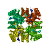
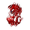
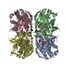
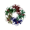
| Title | STRUCTURE OF E. COLI MOBA WITH BOUND GTP AND MANGANESE | ||||||
 Components Components | MOLYBDOPTERIN-GUANINE DINUCLEOTIDE BIOSYNTHESIS PROTEIN | ||||||
 Keywords Keywords | METAL BINDING PROTEIN / Molybdenum cofactor (Moco) / Moco Biosynthesis / Molybdopterin (MPT) / Molybdopterin Guanine Dinucleotide (MGD) | ||||||
| Function / homology |  Function and homology information Function and homology informationbis(molybdopterin guanine dinucleotide)molybdenum biosynthetic process /  molybdenum cofactor guanylyltransferase / molybdenum cofactor guanylyltransferase activity / nucleotidyltransferase activity / GTP binding / magnesium ion binding / cytoplasm molybdenum cofactor guanylyltransferase / molybdenum cofactor guanylyltransferase activity / nucleotidyltransferase activity / GTP binding / magnesium ion binding / cytoplasmSimilarity search - Function | ||||||
| Biological species |  Escherichia coli (E. coli) Escherichia coli (E. coli) | ||||||
| Method | X-RAY DIFFRACTION / SYNCHROTRON / Resolution: 1.75 Å | ||||||
 Authors Authors | Lake, M.W. / Temple, C.A. / Rajagopalan, K.V. / Schindelin, H. | ||||||
 Citation Citation | Journal: J.Biol.Chem. / Year: 2000 Title: The crystal structure of the Escherichia coli MobA protein provides insight into molybdopterin guanine dinucleotide biosynthesis. Authors: Lake, M.W. / Temple, C.A. / Rajagopalan, K.V. / Schindelin, H. | ||||||
| History |
|
- Structure visualization
| Structure viewer | Molecule: MolmilJmol/JSmol |
|---|
- Downloads & links
-Download
| PDBx/mmCIF format | 1frw.cif.gz | 56.1 KB | Display | PDBx/mmCIF format |
|---|---|---|---|---|
| PDB format | pdb1frw.ent.gz | 39.6 KB | Display | PDB format |
| PDBx/mmJSON format | 1frw.json.gz | Tree view | PDBx/mmJSON format | |
| Others |  Other downloads Other downloads |
-Validation report
| Arichive directory | https://data.pdbj.org/pub/pdb/validation_reports/fr/1frwftp://data.pdbj.org/pub/pdb/validation_reports/fr/1frw | HTTPS FTP |
|---|
-Related structure data

-Links
 PDBj
PDBj

- Assembly
| Deposited unit | 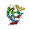
| ||||||||||
|---|---|---|---|---|---|---|---|---|---|---|---|
| 1 | x 8
| ||||||||||
| Unit cell |
| ||||||||||
| Components on special symmetry positions |
| ||||||||||
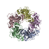
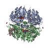
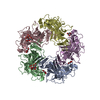
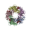
| Details | The biological assembly is an octamer constructed from chain A. The octamer has 42 symmetry and is entirely generated by crystallographic symmetry operations in the I422 tetragonal space group. His 49 from 2 different monomers along with 2 acetate ligands coordinate a zinc atom at the dimer interface. |
-Components
-Protein , 1 types, 1 molecules A
| #1: Protein | Mass: 21669.854 Da / Num. of mol.: 1 / Fragment: MOBA Source method: isolated from a genetically manipulated source Source: (gene. exp.) Escherichia coli (E. coli) / Plasmid: PCT800A / Production host: Escherichia coli (E. coli) / References: UniProt: P32173 |
|---|
-Non-polymers , 5 types, 172 molecules 


| #2: Chemical | ChemComp-ACT / Acetate Mass: 59.044 Da / Num. of mol.: 1 / Source method: obtained synthetically / Formula: C2H3O2 Mass: 59.044 Da / Num. of mol.: 1 / Source method: obtained synthetically / Formula: C2H3O2 |
|---|---|
| #3: Chemical | ChemComp-ZN /  Mass: 65.409 Da / Num. of mol.: 1 / Source method: obtained synthetically / Formula: Zn Mass: 65.409 Da / Num. of mol.: 1 / Source method: obtained synthetically / Formula: Zn |
| #4: Chemical | ChemComp-MN /  Mass: 54.938 Da / Num. of mol.: 1 / Source method: obtained synthetically / Formula: Mn Mass: 54.938 Da / Num. of mol.: 1 / Source method: obtained synthetically / Formula: Mn |
| #5: Chemical | ChemComp-GTP / Guanosine triphosphate Mass: 523.180 Da / Num. of mol.: 1 / Source method: obtained synthetically / Formula: C10H16N5O14P3 / Comment: GTP, energy-carrying molecule*YM Mass: 523.180 Da / Num. of mol.: 1 / Source method: obtained synthetically / Formula: C10H16N5O14P3 / Comment: GTP, energy-carrying molecule*YM |
| #6: Water | ChemComp-HOH / WaterMass: 18.015 Da / Num. of mol.: 168 / Source method: isolated from a natural source / Formula: H2O |
-Experimental details
-Experiment
| Experiment | Method: X-RAY DIFFRACTION / Number of used crystals: 1 |
|---|
- Sample preparation
| Crystal | Density Matthews: 3.01 Å3/Da / Density % sol: 59.08 % | ||||||||||||||||||||||||
|---|---|---|---|---|---|---|---|---|---|---|---|---|---|---|---|---|---|---|---|---|---|---|---|---|---|
| Crystal grow | Temperature: 295 K / Method: vapor diffusion, hanging drop / pH: 6.5 Details: sodium acetate, cacodylate, pH 6.5, VAPOR DIFFUSION, HANGING DROP, temperature 295.0K | ||||||||||||||||||||||||
| Crystal grow | *PLUS Method: vapor diffusion | ||||||||||||||||||||||||
| Components of the solutions | *PLUS
|
-Data collection
| Diffraction | Mean temperature: 95 K |
|---|---|
| Diffraction source | Source: SYNCHROTRON / Site: NSLS  / Beamline: X26C / Wavelength: 1.1 / Beamline: X26C / Wavelength: 1.1 |
| Detector | Type: ADSC QUANTUM 4 / Detector: CCD / Date: Jul 19, 2000 |
| Radiation | Protocol: SINGLE WAVELENGTH / Monochromatic (M) / Laue (L): M / Scattering type: x-ray |
| Radiation wavelength | Wavelength: 1.1 Å / Relative weight: 1 |
| Reflection | Resolution: 1.75→50 Å / Num. all: 26350 / Num. obs: 26350 / % possible obs: 99.6 % / Observed criterion σ(F): 0 / Observed criterion σ(I): 0 / Redundancy: 4.8 % / Biso Wilson estimate: 27.5 Å2 / Rmerge(I) obs: 0.084 / Net I/σ(I): 14 |
| Reflection shell | Resolution: 1.75→1.83 Å / Redundancy: 4.4 % / Rmerge(I) obs: 0.441 / Num. unique all: 2895 / % possible all: 100 |
| Reflection | *PLUS |
| Reflection shell | *PLUS % possible obs: 100 % / Mean I/σ(I) obs: 1.9 |
- Processing
| Software |
| ||||||||||||||||||||||||||||||||||||||||
|---|---|---|---|---|---|---|---|---|---|---|---|---|---|---|---|---|---|---|---|---|---|---|---|---|---|---|---|---|---|---|---|---|---|---|---|---|---|---|---|---|---|
| Refinement | Resolution: 1.75→20 Å / Cross valid method: THROUGHOUT / σ(F): 0 / σ(I): 0 / Stereochemistry target values: Refmac dictionary / Details: Hydrogens have been added in the riding positions
| ||||||||||||||||||||||||||||||||||||||||
| Solvent computation | Solvent model: Babinet's principle | ||||||||||||||||||||||||||||||||||||||||
| Displacement parameters | Biso mean: 22.9 Å2
| ||||||||||||||||||||||||||||||||||||||||
| Refinement step | Cycle: LAST / Resolution: 1.75→20 Å
| ||||||||||||||||||||||||||||||||||||||||
| Refine LS restraints |
| ||||||||||||||||||||||||||||||||||||||||
| Software | *PLUS Name: REFMAC / Classification: refinement | ||||||||||||||||||||||||||||||||||||||||
| Refinement | *PLUS σ(F): 0 / % reflection Rfree: 5.1 % | ||||||||||||||||||||||||||||||||||||||||
| Solvent computation | *PLUS | ||||||||||||||||||||||||||||||||||||||||
| Displacement parameters | *PLUS |
