 Movie
Movie Controller

[English] 日本語
 Yorodumi
- PDB-1fqg: MOLECULAR STRUCTURE OF THE ACYL-ENZYME INTERMEDIATE IN TEM-1 BETA... -
+ Open data

- Basic information
| Entry | Database: PDB / ID: 1fqg | ||||||
|---|---|---|---|---|---|---|---|
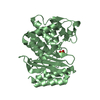
| Title | MOLECULAR STRUCTURE OF THE ACYL-ENZYME INTERMEDIATE IN TEM-1 BETA-LACTAMASE | ||||||
 Components Components | TEM-1 BETA-LACTAMASE | ||||||
 Keywords Keywords |  HYDROLASE / beta-lactamase / acyl-enzyme / penicillin / class A HYDROLASE / beta-lactamase / acyl-enzyme / penicillin / class A | ||||||
| Function / homology |  Function and homology information Function and homology informationbeta-lactam antibiotic catabolic process / beta-lactamase activity / beta-lactamase / response to antibioticSimilarity search - Function | ||||||
| Biological species |  Escherichia coli (E. coli) Escherichia coli (E. coli) | ||||||
| Method | X-RAY DIFFRACTION / SYNCHROTRON / Resolution: 1.7 Å | ||||||
 Authors Authors | Strynadka, N.C. | ||||||
 Citation Citation | Journal: Nature / Year: 1992 Title: Molecular structure of the acyl-enzyme intermediate in beta-lactam hydrolysis at 1.7 A resolution. Authors: Strynadka, N.C. / Adachi, H. / Jensen, S.E. / Johns, K. / Sielecki, A. / Betzel, C. / Sutoh, K. / James, M.N. | ||||||
| History |
|
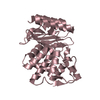
- Structure visualization
| Structure viewer | Molecule: MolmilJmol/JSmol |
|---|
- Downloads & links
-Download
| PDBx/mmCIF format | 1fqg.cif.gz | 62 KB | Display | PDBx/mmCIF format |
|---|---|---|---|---|
| PDB format | pdb1fqg.ent.gz | 48.6 KB | Display | PDB format |
| PDBx/mmJSON format | 1fqg.json.gz | Tree view | PDBx/mmJSON format | |
| Others |  Other downloads Other downloads |
-Validation report
| Arichive directory | https://data.pdbj.org/pub/pdb/validation_reports/fq/1fqgftp://data.pdbj.org/pub/pdb/validation_reports/fq/1fqg | HTTPS FTP |
|---|
-Related structure data
| Related structure data | |
|---|---|
| Similar structure data |


-Links
 PDBj
PDBj


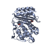
- Assembly
| Deposited unit | 
| ||||||||
|---|---|---|---|---|---|---|---|---|---|
| 1 |
| ||||||||
| Unit cell |
|
-Components
| #1: Protein | Mass: 28926.982 Da / Num. of mol.: 1 / Mutation: GLU166ASN MUTATION Source method: isolated from a genetically manipulated source Source: (gene. exp.) Escherichia coli (E. coli) / Plasmid: PHA508 / Production host: Escherichia coli (E. coli) / References: UniProt: P62593, beta-lactamase |
|---|---|
| #2: Chemical | ChemComp-PNM / Benzylpenicillin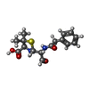  Mass: 336.406 Da / Num. of mol.: 1 / Source method: obtained synthetically / Formula: C16H20N2O4S / Comment: antibiotic*YM Mass: 336.406 Da / Num. of mol.: 1 / Source method: obtained synthetically / Formula: C16H20N2O4S / Comment: antibiotic*YM |
| #3: Water | ChemComp-HOH / Water Mass: 18.015 Da / Num. of mol.: 150 / Source method: isolated from a natural source / Formula: H2O Mass: 18.015 Da / Num. of mol.: 150 / Source method: isolated from a natural source / Formula: H2O |
-Experimental details
-Experiment
| Experiment | Method: X-RAY DIFFRACTION / Number of used crystals: 1 |
|---|
- Sample preparation
| Crystal | Density Matthews: 1.97 Å3/Da / Density % sol: 37.68 % |
|---|---|
| Crystal grow | Temperature: 298 K / Method: vapor diffusion, hanging drop / pH: 8 Details: 1.4M sodium potassium phosphate, pH 8.5, VAPOR DIFFUSION, HANGING DROP, temperature 298K |
| Crystal grow | *PLUS Method: other / Details: used macroseeding |
| Components of the solutions | *PLUS Conc.: 1.4 M / Common name: sodium potassium phosphate / Details: pH8 |
-Data collection
| Diffraction | Mean temperature: 298 K |
|---|---|
| Diffraction source | Source: SYNCHROTRON / Site: EMBL/DESY, HAMBURG  / Beamline: X31 / Wavelength: 1.009 / Beamline: X31 / Wavelength: 1.009 |
| Detector | Type: MARRESEARCH / Detector: IMAGE PLATE / Date: Apr 15, 1992 |
| Radiation | Protocol: SINGLE WAVELENGTH / Monochromatic (M) / Laue (L): M / Scattering type: x-ray |
| Radiation wavelength | Wavelength: 1.009 Å / Relative weight: 1 |
| Reflection | Resolution: 1.7→10 Å / Num. all: 88517 / Num. obs: 24067 / % possible obs: 92 % / Observed criterion σ(F): 2 / Observed criterion σ(I): 1 / Redundancy: 4.2 % / Biso Wilson estimate: 23 Å2 / Rmerge(I) obs: 0.05 / Net I/σ(I): 10.2 |
| Reflection shell | Resolution: 1.7→1.74 Å / Redundancy: 2.2 % / Rmerge(I) obs: 0.106 / Num. unique all: 1622 / % possible all: 82.9 |
| Reflection | *PLUS Rmerge(I) obs: 0.22 |
- Processing
| Software |
| |||||||||||||||||||||||||||||||||||||||||||||
|---|---|---|---|---|---|---|---|---|---|---|---|---|---|---|---|---|---|---|---|---|---|---|---|---|---|---|---|---|---|---|---|---|---|---|---|---|---|---|---|---|---|---|---|---|---|---|
| Refinement | Resolution: 1.7→10 Å / σ(F): 2 / σ(I): 2 / Stereochemistry target values: prolsq
| |||||||||||||||||||||||||||||||||||||||||||||
| Solvent computation | Solvent model: TNT / Bsol: 216.069 Å2 / ksol: 1.13506 e/Å3 | |||||||||||||||||||||||||||||||||||||||||||||
| Refinement step | Cycle: LAST / Resolution: 1.7→10 Å
| |||||||||||||||||||||||||||||||||||||||||||||
| Refine LS restraints |
| |||||||||||||||||||||||||||||||||||||||||||||
| Refinement | *PLUS Lowest resolution: 10 Å | |||||||||||||||||||||||||||||||||||||||||||||
| Solvent computation | *PLUS | |||||||||||||||||||||||||||||||||||||||||||||
| Displacement parameters | *PLUS | |||||||||||||||||||||||||||||||||||||||||||||
| Refine LS restraints | *PLUS
|
