 Movie
Movie Controller

[English] 日本語
 Yorodumi
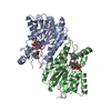
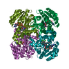
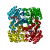
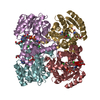
- PDB-1fmc: 7-ALPHA-HYDROXYSTEROID DEHYDROGENASE COMPLEX WITH NADH AND 7-OXO ... -
+ Open data

- Basic information
| Entry | Database: PDB / ID: 1fmc | ||||||
|---|---|---|---|---|---|---|---|
| Title | 7-ALPHA-HYDROXYSTEROID DEHYDROGENASE COMPLEX WITH NADH AND 7-OXO GLYCOCHENODEOXYCHOLIC ACID | ||||||
 Components Components | 7 ALPHA-HYDROXYSTEROID DEHYDROGENASE | ||||||
 Keywords Keywords |  OXIDOREDUCTASE / SHORT-CHAIN DEHYDROGENASE/REDUCTASE / BILE ACID CATABOLISM OXIDOREDUCTASE / SHORT-CHAIN DEHYDROGENASE/REDUCTASE / BILE ACID CATABOLISM | ||||||
| Function / homology |  Function and homology information Function and homology informationchenodeoxycholate 7-alpha-dehydrogenase (NAD+) activity / 7alpha-hydroxysteroid dehydrogenase / cholate 7-alpha-dehydrogenase activity / bile acid catabolic process / lipid catabolic process / NAD binding / protein-containing complex / identical protein binding / cytosolSimilarity search - Function | ||||||
| Biological species |  Escherichia coli (E. coli) Escherichia coli (E. coli) | ||||||
| Method | X-RAY DIFFRACTION / SYNCHROTRON / Resolution: 1.8 Å | ||||||
 Authors Authors | Tanaka, N. / Nonaka, T. / Mitsui, Y. | ||||||
 Citation Citation | Journal: Biochemistry / Year: 1996 Title: Crystal structures of the binary and ternary complexes of 7 alpha-hydroxysteroid dehydrogenase from Escherichia coli. Authors: Tanaka, N. / Nonaka, T. / Tanabe, T. / Yoshimoto, T. / Tsuru, D. / Mitsui, Y. #1: Journal: Acta Crystallogr.,Sect.D / Year: 1996Title: Crystallization and Preliminary X-Ray Crystallographic Studies of 7Alpha-Hydroxysteroid Dehydrogenase from Escherichia Coli Authors: Tanaka, N. / Nonaka, T. / Yoshimoto, T. / Tsuru, D. / Mitsui, Y. #2: Journal: J.Bacteriol. / Year: 1991Title: Cloning and Sequencing of the 7 Alpha-Hydroxysteroid Dehydrogenase Gene from Escherichia Coli Hb101 and Characterization of the Expressed Enzyme Authors: Yoshimoto, T. / Higashi, H. / Kanatani, A. / Lin, X.S. / Nagai, H. / Oyama, H. / Kurazono, K. / Tsuru, D. | ||||||
| History |
|
- Structure visualization
| Structure viewer | Molecule: MolmilJmol/JSmol |
|---|
- Downloads & links
-Download
| PDBx/mmCIF format | 1fmc.cif.gz | 112.5 KB | Display | PDBx/mmCIF format |
|---|---|---|---|---|
| PDB format | pdb1fmc.ent.gz | 87.7 KB | Display | PDB format |
| PDBx/mmJSON format | 1fmc.json.gz | Tree view | PDBx/mmJSON format | |
| Others |  Other downloads Other downloads |
-Validation report
| Arichive directory | https://data.pdbj.org/pub/pdb/validation_reports/fm/1fmcftp://data.pdbj.org/pub/pdb/validation_reports/fm/1fmc | HTTPS FTP |
|---|
-Related structure data


-Links
 PDBj
PDBj


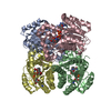
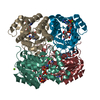
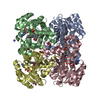
- Assembly
| Deposited unit | 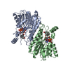
| ||||||||
|---|---|---|---|---|---|---|---|---|---|
| 1 | 
| ||||||||
| Unit cell |
| ||||||||
| Noncrystallographic symmetry (NCS) | NCS oper: (Code: given Matrix: (-0.15963, -0.842545, -0.514428), Vector : |
-Components
| #1: Protein | Mass: 26801.527 Da / Num. of mol.: 2 Source method: isolated from a genetically manipulated source Details: LIGANDS ARE NADH AND 7-OXO GLYCOCHENODEOXYCHOLIC ACID (CHO) Source: (gene. exp.) Escherichia coli (E. coli) / Strain: HB101 / Description: COLON BACILLUS / Cell line: CHO / Plasmid: HB101 / Production host: Escherichia coli (E. coli)References: UniProt: P25529, UniProt: P0AET8*PLUS, 7alpha-hydroxysteroid dehydrogenase#2: Chemical | Glycochenodeoxycholic acid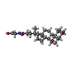  Mass: 449.623 Da / Num. of mol.: 2 / Source method: obtained synthetically / Formula: C26H43NO5 / Comment: detergent*YM Mass: 449.623 Da / Num. of mol.: 2 / Source method: obtained synthetically / Formula: C26H43NO5 / Comment: detergent*YM#3: Chemical | Nicotinamide adenine dinucleotide  Mass: 665.441 Da / Num. of mol.: 2 / Source method: obtained synthetically / Formula: C21H29N7O14P2 Mass: 665.441 Da / Num. of mol.: 2 / Source method: obtained synthetically / Formula: C21H29N7O14P2#4: Water | ChemComp-HOH / | Water Mass: 18.015 Da / Num. of mol.: 242 / Source method: isolated from a natural source / Formula: H2O Mass: 18.015 Da / Num. of mol.: 242 / Source method: isolated from a natural source / Formula: H2O |
|---|
-Experimental details
-Experiment
| Experiment | Method: X-RAY DIFFRACTION |
|---|
- Sample preparation
| Crystal | Density Matthews: 3.34 Å3/Da / Density % sol: 62 % | ||||||||||||||||||||||||||||||
|---|---|---|---|---|---|---|---|---|---|---|---|---|---|---|---|---|---|---|---|---|---|---|---|---|---|---|---|---|---|---|---|
| Crystal grow | pH: 8.5 / Details: pH 8.5 | ||||||||||||||||||||||||||||||
| Crystal grow | *PLUS Temperature: 293 K / Method: vapor diffusion, hanging dropDetails: drop solution was prepared by mixing the protein, reservoir and detergent solutions, buffered by 100mM Tris, at the ratio of 4:3:1. | ||||||||||||||||||||||||||||||
| Components of the solutions | *PLUS
|
-Data collection
| Diffraction | Mean temperature: 293 K |
|---|---|
| Diffraction source | Source: SYNCHROTRON / Site: Photon Factory  / Beamline: BL-18B / Wavelength: 1 / Beamline: BL-18B / Wavelength: 1 |
| Detector | Type: WEISSENBERG / Detector: DIFFRACTOMETER / Date: Apr 23, 1995 / Details: BENT CYLINDER |
| Radiation | Monochromator: DOUBLE CRYSTAL SI(111) / Monochromatic (M) / Laue (L): M / Scattering type: x-ray |
| Radiation wavelength | Wavelength: 1 Å / Relative weight: 1 |
| Reflection | Resolution: 1.8→76.7 Å / Num. obs: 57290 / % possible obs: 84 % / Observed criterion σ(I): 1 / Redundancy: 3.9 % / Rmerge(I) obs: 0.085 |
| Reflection | *PLUS Num. measured all: 221323 |
| Reflection shell | *PLUS % possible obs: 66.1 % / Rmerge(I) obs: 0.397 |
- Processing
| Software |
| ||||||||||||||||||||||||||||||||||||||||||||||||||||||||||||
|---|---|---|---|---|---|---|---|---|---|---|---|---|---|---|---|---|---|---|---|---|---|---|---|---|---|---|---|---|---|---|---|---|---|---|---|---|---|---|---|---|---|---|---|---|---|---|---|---|---|---|---|---|---|---|---|---|---|---|---|---|---|
| Refinement | Resolution: 1.8→8 Å / σ(F): 1
| ||||||||||||||||||||||||||||||||||||||||||||||||||||||||||||
| Displacement parameters | Biso mean: 26.1 Å2 | ||||||||||||||||||||||||||||||||||||||||||||||||||||||||||||
| Refine analyze | Luzzati coordinate error obs: 0.25 Å | ||||||||||||||||||||||||||||||||||||||||||||||||||||||||||||
| Refinement step | Cycle: LAST / Resolution: 1.8→8 Å
| ||||||||||||||||||||||||||||||||||||||||||||||||||||||||||||
| Refine LS restraints |
| ||||||||||||||||||||||||||||||||||||||||||||||||||||||||||||
| Software | *PLUS Name: X-PLOR / Classification: refinement | ||||||||||||||||||||||||||||||||||||||||||||||||||||||||||||
| Refinement | *PLUS Rfactor all: 0.252 | ||||||||||||||||||||||||||||||||||||||||||||||||||||||||||||
| Solvent computation | *PLUS | ||||||||||||||||||||||||||||||||||||||||||||||||||||||||||||
| Displacement parameters | *PLUS | ||||||||||||||||||||||||||||||||||||||||||||||||||||||||||||
| Refine LS restraints | *PLUS
|
