 Movie
Movie Controller

[English] 日本語
 Yorodumi
- PDB-1fda: CRYSTAL STRUCTURES OF OXIDIZED AND REDUCED AZOTOBACTER VINELANDII... -
+ Open data

- Basic information
| Entry | Database: PDB / ID: 1fda | ||||||
|---|---|---|---|---|---|---|---|
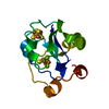
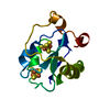
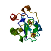
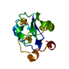
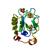
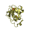
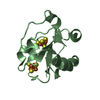
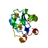
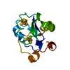
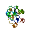
| Title | CRYSTAL STRUCTURES OF OXIDIZED AND REDUCED AZOTOBACTER VINELANDII FERREDOXIN AT PH 8 AND PH 6 | ||||||
 Components Components | FERREDOXIN | ||||||
 Keywords Keywords | ELECTRON TRANSPORT(IRON-SULFUR) | ||||||
| Function / homology |  Function and homology information Function and homology information3 iron, 4 sulfur cluster binding / 4 iron, 4 sulfur cluster binding / electron transfer activity / DNA binding / metal ion bindingSimilarity search - Function | ||||||
| Biological species |  Azotobacter vinelandii (bacteria) Azotobacter vinelandii (bacteria) | ||||||
| Method | X-RAY DIFFRACTION / Resolution: 2.1 Å | ||||||
 Authors Authors | Stout, C.D. | ||||||
 Citation Citation | Journal: J.Biol.Chem. / Year: 1993 Title: Crystal structures of oxidized and reduced Azotobacter vinelandii ferredoxin at pH 8 and 6. Authors: Stout, C.D. #1: Journal: J.Biol.Chem. / Year: 1991Title: Crystallographic Analysis of Two Site-Directed Mutants of Azotobacter Vinelandii Ferredoxin Authors: Soman, J. / Iismaa, S. / Stout, C.D. #2: Journal: Proc.Natl.Acad.Sci.USA / Year: 1990Title: Site-Directed Mutagenesis of Azotobacter Vinelandii Ferredoxin I. (Fe-S) Cluster-Driven Protein Rearrangement Authors: Martin, A.E. / Burgess, B.K. / Stout, C.D. / Cash, V.L. / Dean, D.R. / Jensen, G.M. / Stephens, P.J. #3: Journal: J.Mol.Biol. / Year: 1989Title: Refinement of the 7 Fe Ferredoxin from Azotobacter at 1.9 Angstroms Resolution Authors: Stout, C.D. #5: Journal: Proc.Natl.Acad.Sci.USA / Year: 1985Title: (4Fe-4S)-Cluster-Depleted Azotobacter Vinelandii Ferredoxin I. A New 3Fe Iron-Sulfur Protein Authors: Stephens, P.J. / Morgan, T.V. / Devlin, F. / Penner-Hahn, J.E. / Hodgson, K.O. / Scott, R.A. / Stout, C.D. / Burgess, B.K. #6: Journal: J.Biol.Chem. / Year: 1983Title: Structure of Azotobacter Vinelandii 7Fe Ferredoxin. Amino Acid Sequence and Electron Density Maps of Residues Authors: Howard, J.B. / Lorsbach, T.W. / Ghosh, D. / Melis, K. / Stout, C.D. #7: Journal: J.Mol.Biol. / Year: 1982Title: Iron-Sulfur Clusters and Protein Structure of Azotobacter Ferredoxin at 2.0 Angstroms Resolution Authors: Ghosh, D. / O'Donnell, S. / Fureyjunior, W. / Robbins, A.H. / Stout, C.D. #8: Journal: J.Biol.Chem. / Year: 1981Title: Structure of a 7Fe Ferredoxin from Azotobacter Vinelandii Authors: Ghosh, D. / Fureyjunior, W. / O'Donnell, S. / Stout, C.D. #9: Journal: J.Biol.Chem. / Year: 1980Title: Iron-Sulfur Clusters in Azotobacter Ferredoxin at 2.5 Angstroms Resolution Authors: Stout, C.D. / Ghosh, D. / Pattabhi, V. / Robbins, A.H. #10: Journal: Am.Cryst.Assoc.,Abstr.Papers (Winter Meeting) / Year: 1979Title: Structure of the Iron-Sulfur Clusters in Azotobacter Ferredoxin at 4.0 Angstroms Resolution Authors: Stout, C.D. #11: Journal: J.Biol.Chem. / Year: 1979Title: Two Crystal Forms of Azotobacter Ferredoxin Authors: Stout, C.D. #12: Journal: Nature / Year: 1979Title: Structure of the Iron-Sulphur Clusters in Azotobacter Ferredoxin at 4.0 Angstroms Resolution Authors: Stout, C.D. | ||||||
| History |
|
- Structure visualization
| Structure viewer | Molecule: MolmilJmol/JSmol |
|---|
- Downloads & links
-Download
| PDBx/mmCIF format | 1fda.cif.gz | 34.8 KB | Display | PDBx/mmCIF format |
|---|---|---|---|---|
| PDB format | pdb1fda.ent.gz | 23.4 KB | Display | PDB format |
| PDBx/mmJSON format | 1fda.json.gz | Tree view | PDBx/mmJSON format | |
| Others |  Other downloads Other downloads |
-Validation report
| Arichive directory | https://data.pdbj.org/pub/pdb/validation_reports/fd/1fdaftp://data.pdbj.org/pub/pdb/validation_reports/fd/1fda | HTTPS FTP |
|---|
-Related structure data


-Links
 PDBj
PDBj


- Assembly
| Deposited unit | 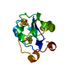
| ||||||||
|---|---|---|---|---|---|---|---|---|---|
| 1 |
| ||||||||
| Unit cell |
|
-Components
| #1: Protein | Mass: 12059.530 Da / Num. of mol.: 1 Source method: isolated from a genetically manipulated source Source: (gene. exp.) Azotobacter vinelandii (bacteria) / References: UniProt: P00214 |
|---|---|
| #2: Chemical | ChemComp-SF4 / Iron–sulfur cluster  Mass: 351.640 Da / Num. of mol.: 1 / Source method: obtained synthetically / Formula: Fe4S4 Mass: 351.640 Da / Num. of mol.: 1 / Source method: obtained synthetically / Formula: Fe4S4 |
| #3: Chemical | ChemComp-F3S / Iron–sulfur cluster  Mass: 295.795 Da / Num. of mol.: 1 / Source method: obtained synthetically / Formula: Fe3S4 Mass: 295.795 Da / Num. of mol.: 1 / Source method: obtained synthetically / Formula: Fe3S4 |
| #4: Water | ChemComp-HOH / Water Mass: 18.015 Da / Num. of mol.: 36 / Source method: isolated from a natural source / Formula: H2O Mass: 18.015 Da / Num. of mol.: 36 / Source method: isolated from a natural source / Formula: H2O |
| Sequence details | THE CHEMICAL SEQUENCE WAS DETERMINED IN COLLABORATION WITH J. B. HOWARD AND T. LORSBACH OF DEPT. OF ...THE CHEMICAL SEQUENCE WAS DETERMINED |
-Experimental details
-Experiment
| Experiment | Method: X-RAY DIFFRACTION |
|---|
- Sample preparation
| Crystal | Density Matthews: 3 Å3/Da / Density % sol: 59.02 % | |||||||||||||||||||||||||
|---|---|---|---|---|---|---|---|---|---|---|---|---|---|---|---|---|---|---|---|---|---|---|---|---|---|---|
| Crystal | *PLUS Density % sol: 55 % | |||||||||||||||||||||||||
| Crystal grow | *PLUS Temperature: 2 ℃ / pH: 7.4 / Method: vapor diffusion / Details: referred to J.Biol.Chem. 254.3598-3599 1979 | |||||||||||||||||||||||||
| Components of the solutions | *PLUS
|
-Data collection
| Radiation | Scattering type: x-ray |
|---|---|
| Radiation wavelength | Relative weight: 1 |
| Reflection | *PLUS Highest resolution: 2.1 Å / Lowest resolution: 20 Å / Num. all: 9115 / Num. obs: 8487 / % possible obs: 93.1 % / Num. measured all: 36781 / Rmerge(I) obs: 0.062 |
- Processing
| Software |
| ||||||||||||||||||||||||||||||||||||||||||||||||||||||||||||
|---|---|---|---|---|---|---|---|---|---|---|---|---|---|---|---|---|---|---|---|---|---|---|---|---|---|---|---|---|---|---|---|---|---|---|---|---|---|---|---|---|---|---|---|---|---|---|---|---|---|---|---|---|---|---|---|---|---|---|---|---|---|
| Refinement | Resolution: 2.1→8 Å / σ(F): 0 /
| ||||||||||||||||||||||||||||||||||||||||||||||||||||||||||||
| Refinement step | Cycle: LAST / Resolution: 2.1→8 Å
| ||||||||||||||||||||||||||||||||||||||||||||||||||||||||||||
| Refine LS restraints |
| ||||||||||||||||||||||||||||||||||||||||||||||||||||||||||||
| Refinement | *PLUS Highest resolution: 2.1 Å / Lowest resolution: 8 Å / Num. reflection all: 7981 / σ(F): 0 / Rfactor all: 0.206 | ||||||||||||||||||||||||||||||||||||||||||||||||||||||||||||
| Solvent computation | *PLUS | ||||||||||||||||||||||||||||||||||||||||||||||||||||||||||||
| Displacement parameters | *PLUS Biso mean: 17.2 Å2 | ||||||||||||||||||||||||||||||||||||||||||||||||||||||||||||
| Refine LS restraints | *PLUS
|
