 Movie
Movie Controller

[English] 日本語
 Yorodumi
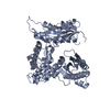
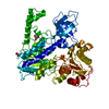
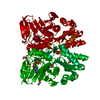
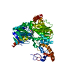
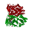
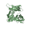
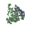
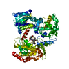
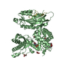
- PDB-1fa0: STRUCTURE OF YEAST POLY(A) POLYMERASE BOUND TO MANGANATE AND 3'-DATP -
+ Open data

- Basic information
| Entry | Database: PDB / ID: 1fa0 | ||||||
|---|---|---|---|---|---|---|---|
| Title | STRUCTURE OF YEAST POLY(A) POLYMERASE BOUND TO MANGANATE AND 3'-DATP | ||||||
 Components Components | POLY(A)-POLYMERASE | ||||||
 Keywords Keywords |  TRANSFERASE / Polymerase / Nucleotidyl Transferase TRANSFERASE / Polymerase / Nucleotidyl Transferase | ||||||
| Function / homology |  Function and homology information Function and homology informationtermination of RNA polymerase II transcription, poly(A)-coupled / sno(s)RNA 3'-end processing / mRNA cleavage and polyadenylation specificity factor complex / polynucleotide adenylyltransferase / poly(A) RNA polymerase activity / mRNA 3'-end processing / : / magnesium ion binding / RNA binding / ATP binding / nucleusSimilarity search - Function | ||||||
| Biological species |  Saccharomyces cerevisiae (brewer's yeast) Saccharomyces cerevisiae (brewer's yeast) | ||||||
| Method | X-RAY DIFFRACTION / SYNCHROTRON / Resolution: 2.6 Å | ||||||
 Authors Authors | Bard, J. / Zhelkovsky, A.M. / Helmling, S. / Moore, C.L. / Bohm, A. | ||||||
 Citation Citation | Journal: Science / Year: 2000 Title: Structure of yeast poly(A) polymerase alone and in complex with 3'-dATP. Authors: Bard, J. / Zhelkovsky, A.M. / Helmling, S. / Earnest, T.N. / Moore, C.L. / Bohm, A. | ||||||
| History |
|
- Structure visualization
| Structure viewer | Molecule: MolmilJmol/JSmol |
|---|
- Downloads & links
-Download
| PDBx/mmCIF format | 1fa0.cif.gz | 213.9 KB | Display | PDBx/mmCIF format |
|---|---|---|---|---|
| PDB format | pdb1fa0.ent.gz | 170.6 KB | Display | PDB format |
| PDBx/mmJSON format | 1fa0.json.gz | Tree view | PDBx/mmJSON format | |
| Others |  Other downloads Other downloads |
-Validation report
| Arichive directory | https://data.pdbj.org/pub/pdb/validation_reports/fa/1fa0ftp://data.pdbj.org/pub/pdb/validation_reports/fa/1fa0 | HTTPS FTP |
|---|
-Related structure data
| Similar structure data |
|---|

-Links
 PDBj
PDBj


- Assembly
| Deposited unit | 
| ||||||||
|---|---|---|---|---|---|---|---|---|---|
| 1 | 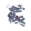
| ||||||||
| 2 | 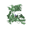
| ||||||||
| Unit cell |
|
-Components
-Protein , 1 types, 2 molecules AB
| #1: Protein | Mass: 61439.508 Da / Num. of mol.: 2 / Fragment: RESIDUES 1-530 Source method: isolated from a genetically manipulated source Source: (gene. exp.) Saccharomyces cerevisiae (brewer's yeast)Plasmid: PJDELTA10PAP / Production host:  Escherichia coli (E. coli) Escherichia coli (E. coli)References: UniProt: P29468, polynucleotide adenylyltransferase |
|---|
-Non-polymers , 5 types, 91 molecules 
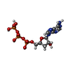
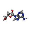


| #2: Chemical | ChemComp-MN /  Mass: 54.938 Da / Num. of mol.: 4 / Source method: obtained synthetically / Formula: Mn Mass: 54.938 Da / Num. of mol.: 4 / Source method: obtained synthetically / Formula: Mn#3: Chemical | Deoxyadenosine triphosphate Mass: 491.182 Da / Num. of mol.: 2 / Source method: obtained synthetically / Formula: C10H16N5O12P3 Mass: 491.182 Da / Num. of mol.: 2 / Source method: obtained synthetically / Formula: C10H16N5O12P3#4: Chemical | Cordycepin Mass: 251.242 Da / Num. of mol.: 2 / Source method: obtained synthetically / Formula: C10H13N5O3 Mass: 251.242 Da / Num. of mol.: 2 / Source method: obtained synthetically / Formula: C10H13N5O3#5: Chemical | ChemComp-POP / | Pyrophosphate Mass: 175.959 Da / Num. of mol.: 1 / Source method: obtained synthetically / Formula: H2O7P2 Mass: 175.959 Da / Num. of mol.: 1 / Source method: obtained synthetically / Formula: H2O7P2#6: Water | ChemComp-HOH / | WaterMass: 18.015 Da / Num. of mol.: 82 / Source method: isolated from a natural source / Formula: H2O |
|---|
-Experimental details
-Experiment
| Experiment | Method: X-RAY DIFFRACTION / Number of used crystals: 1 |
|---|
- Sample preparation
| Crystal | Density Matthews: 3.91 Å3/Da / Density % sol: 68.5 % | |||||||||||||||||||||||||||||||||||
|---|---|---|---|---|---|---|---|---|---|---|---|---|---|---|---|---|---|---|---|---|---|---|---|---|---|---|---|---|---|---|---|---|---|---|---|---|
| Crystal grow | Temperature: 277 K / Method: vapor diffusion, hanging drop / pH: 7.24 Details: PEG,Ethylene Glycol, pH 7.24, VAPOR DIFFUSION, HANGING DROP, temperature 277K | |||||||||||||||||||||||||||||||||||
| Crystal grow | *PLUS Temperature: 4 ℃ | |||||||||||||||||||||||||||||||||||
| Components of the solutions | *PLUS
|
-Data collection
| Diffraction | Mean temperature: 100 K |
|---|---|
| Diffraction source | Source: SYNCHROTRON / Site: APS  / Beamline: 19-ID / Wavelength: 1.033 / Beamline: 19-ID / Wavelength: 1.033 |
| Detector | Type: OTHER / Detector: CCD / Date: Feb 4, 2000 |
| Radiation | Monochromator: MIRRORS / Protocol: SINGLE WAVELENGTH / Monochromatic (M) / Laue (L): M / Scattering type: x-ray |
| Radiation wavelength | Wavelength: 1.033 Å / Relative weight: 1 |
| Reflection | Resolution: 2.6→30 Å / Num. all: 59680 / Num. obs: 58367 / % possible obs: 97.8 % / Observed criterion σ(I): 0 / Redundancy: 4.14 % / Biso Wilson estimate: 44.5 Å2 / Rmerge(I) obs: 0.044 / Net I/σ(I): 32.36 |
| Reflection shell | Resolution: 2.6→2.72 Å / Redundancy: 4.2 % / Rmerge(I) obs: 0.307 / Num. unique all: 7379 / % possible all: 99.9 |
| Reflection | *PLUS Num. measured all: 242159 |
| Reflection shell | *PLUS % possible obs: 99.9 % / Mean I/σ(I) obs: 2.8 |
- Processing
| Software |
| ||||||||||||||||||||||||||||||||||||||||
|---|---|---|---|---|---|---|---|---|---|---|---|---|---|---|---|---|---|---|---|---|---|---|---|---|---|---|---|---|---|---|---|---|---|---|---|---|---|---|---|---|---|
| Refinement | Resolution: 2.6→20 Å / Rfactor Rfree error: 0.006 / Data cutoff high absF: 1052123.24 / Data cutoff low absF: 0 / Isotropic thermal model: RESTRAINED / Cross valid method: THROUGHOUT / σ(F): 2 / Stereochemistry target values: Engh & Huber
| ||||||||||||||||||||||||||||||||||||||||
| Solvent computation | Solvent model: FLAT MODEL / Bsol: 40.7 Å2 / ksol: 0.396 e/Å3 | ||||||||||||||||||||||||||||||||||||||||
| Displacement parameters | Biso mean: 62.7 Å2
| ||||||||||||||||||||||||||||||||||||||||
| Refine analyze |
| ||||||||||||||||||||||||||||||||||||||||
| Refinement step | Cycle: LAST / Resolution: 2.6→20 Å
| ||||||||||||||||||||||||||||||||||||||||
| Refine LS restraints |
| ||||||||||||||||||||||||||||||||||||||||
| LS refinement shell | Resolution: 2.6→2.69 Å / Rfactor Rfree error: 0.017 / Total num. of bins used: 6
| ||||||||||||||||||||||||||||||||||||||||
| Xplor file |
| ||||||||||||||||||||||||||||||||||||||||
| Software | *PLUS Name: CNS / Version: 0.9 / Classification: refinement | ||||||||||||||||||||||||||||||||||||||||
| Refinement | *PLUS Highest resolution: 2.6 Å / Lowest resolution: 20 Å / σ(F): 2 / % reflection Rfree: 8.5 % | ||||||||||||||||||||||||||||||||||||||||
| Solvent computation | *PLUS | ||||||||||||||||||||||||||||||||||||||||
| Displacement parameters | *PLUS Biso mean: 62.7 Å2 | ||||||||||||||||||||||||||||||||||||||||
| Refine LS restraints | *PLUS
| ||||||||||||||||||||||||||||||||||||||||
| LS refinement shell | *PLUS % reflection Rfree: 9.7 % |
