 Movie
Movie Controller

[English] 日本語
 Yorodumi
- PDB-1f12: L-3-HYDROXYACYL-COA DEHYDROGENASE COMPLEXED WITH 3-HYDROXYBUTYRYL-COA -
+ Open data

- Basic information
| Entry | Database: PDB / ID: 1f12 | ||||||
|---|---|---|---|---|---|---|---|
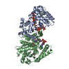
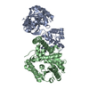
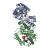
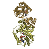
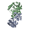
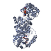
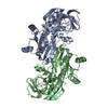
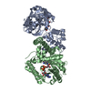
| Title | L-3-HYDROXYACYL-COA DEHYDROGENASE COMPLEXED WITH 3-HYDROXYBUTYRYL-COA | ||||||
 Components Components | L-3-HYDROXYACYL-COA DEHYDROGENASE | ||||||
 Keywords Keywords |  OXIDOREDUCTASE / L-3-hydroxyacyl-CoA complexed with 3-hydroxybutyryl-CoA OXIDOREDUCTASE / L-3-hydroxyacyl-CoA complexed with 3-hydroxybutyryl-CoA | ||||||
| Function / homology |  Function and homology information Function and homology informationBeta oxidation of lauroyl-CoA to decanoyl-CoA-CoA / Beta oxidation of butanoyl-CoA to acetyl-CoA / Beta oxidation of hexanoyl-CoA to butanoyl-CoA / Beta oxidation of octanoyl-CoA to hexanoyl-CoA / Beta oxidation of decanoyl-CoA to octanoyl-CoA-CoA / 3-hydroxyacyl-CoA dehydrogenase / 3-hydroxyacyl-CoA dehydrogenase activity / fatty acid beta-oxidation / regulation of insulin secretion / NAD+ binding ...Beta oxidation of lauroyl-CoA to decanoyl-CoA-CoA / Beta oxidation of butanoyl-CoA to acetyl-CoA / Beta oxidation of hexanoyl-CoA to butanoyl-CoA / Beta oxidation of octanoyl-CoA to hexanoyl-CoA / Beta oxidation of decanoyl-CoA to octanoyl-CoA-CoA / 3-hydroxyacyl-CoA dehydrogenase / 3-hydroxyacyl-CoA dehydrogenase activity / fatty acid beta-oxidation / regulation of insulin secretion / NAD+ binding / negative regulation of insulin secretion / response to activity / response to insulin / positive regulation of cold-induced thermogenesis / transferase activity / mitochondrial matrix / response to xenobiotic stimulus / mitochondrion / nucleoplasm / identical protein binding / cytoplasmSimilarity search - Function | ||||||
| Biological species |  Homo sapiens (human) Homo sapiens (human) | ||||||
| Method | X-RAY DIFFRACTION / SYNCHROTRON / Resolution: 2.4 Å | ||||||
 Authors Authors | Barycki, J.J. / O'Brien, L.K. / Strauss, A.W. / Banaszak, L.J. | ||||||
 Citation Citation | Journal: J.Biol.Chem. / Year: 2000 Title: Sequestration of the active site by interdomain shifting. Crystallographic and spectroscopic evidence for distinct conformations of L-3-hydroxyacyl-CoA dehydrogenase. Authors: Barycki, J.J. / O'Brien, L.K. / Strauss, A.W. / Banaszak, L.J. #1: Journal: Biochemistry / Year: 1999Title: Biochemical Characterization and Crystal Structure Determination of Human Heart Short Chain L-3-hydroxyacyl-CoA Dehydrogenase Provide Insights into Catalytic Mechanism Authors: Barycki, J.J. / O'Brien, L.K. / Bratt, J.M. / Zhang, R. / Sanishvili, R. / Strauss, A.W. / Banaszak, L.J. | ||||||
| History |
|
- Structure visualization
| Structure viewer | Molecule: MolmilJmol/JSmol |
|---|
- Downloads & links
-Download
| PDBx/mmCIF format | 1f12.cif.gz | 125.4 KB | Display | PDBx/mmCIF format |
|---|---|---|---|---|
| PDB format | pdb1f12.ent.gz | 99.1 KB | Display | PDB format |
| PDBx/mmJSON format | 1f12.json.gz | Tree view | PDBx/mmJSON format | |
| Others |  Other downloads Other downloads |
-Validation report
| Arichive directory | https://data.pdbj.org/pub/pdb/validation_reports/f1/1f12ftp://data.pdbj.org/pub/pdb/validation_reports/f1/1f12 | HTTPS FTP |
|---|
-Related structure data


-Links
 PDBj
PDBj


- Assembly
| Deposited unit | 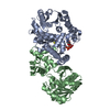
| ||||||||
|---|---|---|---|---|---|---|---|---|---|
| 1 |
| ||||||||
| Unit cell |
|
-Components
| #1: Protein | Mass: 33895.824 Da / Num. of mol.: 2 / Mutation: F80C Source method: isolated from a genetically manipulated source Source: (gene. exp.) Homo sapiens (human)Description: PROTEIN WAS EXPRESSED WITH A C-TERMINAL HEXAMERIC HISTIDINE TAG. Organ: HEART / Plasmid: PET28 / Production host:  Escherichia coli (E. coli) Escherichia coli (E. coli)References: UniProt: Q16836, 3-hydroxyacyl-CoA dehydrogenase#2: Chemical | ChemComp-3HC / |   Mass: 853.623 Da / Num. of mol.: 1 / Source method: obtained synthetically / Formula: C25H42N7O18P3S Mass: 853.623 Da / Num. of mol.: 1 / Source method: obtained synthetically / Formula: C25H42N7O18P3S#3: Water | ChemComp-HOH / | Water Mass: 18.015 Da / Num. of mol.: 187 / Source method: isolated from a natural source / Formula: H2O Mass: 18.015 Da / Num. of mol.: 187 / Source method: isolated from a natural source / Formula: H2O |
|---|
-Experimental details
-Experiment
| Experiment | Method: X-RAY DIFFRACTION / Number of used crystals: 1 |
|---|
- Sample preparation
| Crystal | Density Matthews: 2.72 Å3/Da / Density % sol: 54.74 % | |||||||||||||||||||||||||
|---|---|---|---|---|---|---|---|---|---|---|---|---|---|---|---|---|---|---|---|---|---|---|---|---|---|---|
| Crystal grow | Temperature: 291 K / Method: vapor diffusion, hanging drop / pH: 6.5 Details: 50 mM N-[2-acetamido]-2-iminodiacetic acid within the precipitant range of 14% to 19% polyethylene glycol 4000, pH 6.5, VAPOR DIFFUSION, HANGING DROP, temperature 291K | |||||||||||||||||||||||||
| Crystal grow | *PLUS Details: Barycki, J.J., (1999) Biochemistry, 38, 5786. | |||||||||||||||||||||||||
| Components of the solutions | *PLUS
|
-Data collection
| Diffraction | Mean temperature: 100 K |
|---|---|
| Diffraction source | Source: SYNCHROTRON / Site: APS  / Beamline: 19-ID / Wavelength: 1.03321 / Beamline: 19-ID / Wavelength: 1.03321 |
| Detector | Type: APS-1 / Detector: CCD / Date: Feb 12, 1999 |
| Radiation | Protocol: SINGLE WAVELENGTH / Monochromatic (M) / Laue (L): M / Scattering type: x-ray |
| Radiation wavelength | Wavelength: 1.03321 Å / Relative weight: 1 |
| Reflection | Resolution: 2.4→20 Å / Num. all: 29550 / Num. obs: 29550 / % possible obs: 99.7 % / Observed criterion σ(F): 0 / Observed criterion σ(I): 0 / Redundancy: 4.8 % / Biso Wilson estimate: 49.8 Å2 / Rmerge(I) obs: 0.067 / Net I/σ(I): 20.4 |
| Reflection shell | Resolution: 2.4→2.49 Å / Rmerge(I) obs: 0.474 / Num. unique all: 2908 / % possible all: 99.7 |
| Reflection shell | *PLUS % possible obs: 99.7 % |
- Processing
| Software |
| ||||||||||||||||||||||||||||||||||||||||||||||||||||||||||||||||||||||||||||||||
|---|---|---|---|---|---|---|---|---|---|---|---|---|---|---|---|---|---|---|---|---|---|---|---|---|---|---|---|---|---|---|---|---|---|---|---|---|---|---|---|---|---|---|---|---|---|---|---|---|---|---|---|---|---|---|---|---|---|---|---|---|---|---|---|---|---|---|---|---|---|---|---|---|---|---|---|---|---|---|---|---|---|
| Refinement | Resolution: 2.4→20 Å / Rfactor Rfree error: 0.007 / Data cutoff high absF: 689257.88 / Data cutoff low absF: 0 / Isotropic thermal model: RESTRAINED / Cross valid method: THROUGHOUT / σ(F): 0 / σ(I): 0 / Stereochemistry target values: Engh & Huber
| ||||||||||||||||||||||||||||||||||||||||||||||||||||||||||||||||||||||||||||||||
| Displacement parameters | Biso mean: 42 Å2
| ||||||||||||||||||||||||||||||||||||||||||||||||||||||||||||||||||||||||||||||||
| Refine analyze |
| ||||||||||||||||||||||||||||||||||||||||||||||||||||||||||||||||||||||||||||||||
| Refinement step | Cycle: LAST / Resolution: 2.4→20 Å
| ||||||||||||||||||||||||||||||||||||||||||||||||||||||||||||||||||||||||||||||||
| Refine LS restraints |
| ||||||||||||||||||||||||||||||||||||||||||||||||||||||||||||||||||||||||||||||||
| LS refinement shell | Resolution: 2.4→2.55 Å / Rfactor Rfree error: 0.024 / Total num. of bins used: 6
| ||||||||||||||||||||||||||||||||||||||||||||||||||||||||||||||||||||||||||||||||
| Xplor file |
| ||||||||||||||||||||||||||||||||||||||||||||||||||||||||||||||||||||||||||||||||
| Software | *PLUS Name: CNS / Version: 0.3 / Classification: refinement | ||||||||||||||||||||||||||||||||||||||||||||||||||||||||||||||||||||||||||||||||
| Refine LS restraints | *PLUS
|
