 Movie
Movie Controller

[English] 日本語
 Yorodumi
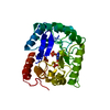
- PDB-1eye: 1.7 ANGSTROM RESOLUTION CRYSTAL STRUCTURE OF 6-HYDROXYMETHYL-7,8-... -
+ Open data

- Basic information
| Entry | Database: PDB / ID: 1eye | ||||||
|---|---|---|---|---|---|---|---|
| Title | 1.7 ANGSTROM RESOLUTION CRYSTAL STRUCTURE OF 6-HYDROXYMETHYL-7,8-DIHYDROPTEROATE SYNTHASE (DHPS) FROM MYCOBACTERIUM TUBERCULOSIS IN COMPLEX WITH 6-HYDROXYMETHYLPTERIN MONOPHOSPHATE | ||||||
 Components Components | DIHYDROPTEROATE SYNTHASE I | ||||||
 Keywords Keywords | TRANSFERASE / alpha-beta barrel | ||||||
| Function / homology |  Function and homology informationdihydropteroate synthase / dihydropteroate synthase activity / folic acid biosynthetic process / tetrahydrofolate biosynthetic process / metal ion binding / cytosol Function and homology informationdihydropteroate synthase / dihydropteroate synthase activity / folic acid biosynthetic process / tetrahydrofolate biosynthetic process / metal ion binding / cytosolSimilarity search - Function | ||||||
| Biological species |  Mycobacterium tuberculosis H37Rv (bacteria) Mycobacterium tuberculosis H37Rv (bacteria) | ||||||
| Method | X-RAY DIFFRACTION / Resolution: 1.7 Å | ||||||
 Authors Authors | Baca, A.M. / Sirawaraporn, R. / Turley, S. / Sirawaraporn, W. / Hol, W.G.J. | ||||||
 Citation Citation | Journal: J.Mol.Biol. / Year: 2000 Title: Crystal structure of Mycobacterium tuberculosis 7,8-dihydropteroate synthase in complex with pterin monophosphate: new insight into the enzymatic mechanism and sulfa-drug action. Authors: Baca, A.M. / Sirawaraporn, R. / Turley, S. / Sirawaraporn, W. / Hol, W.G. | ||||||
| History |
|
- Structure visualization
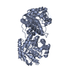
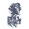
| Structure viewer | Molecule: MolmilJmol/JSmol |
|---|
- Downloads & links
-Download
| PDBx/mmCIF format | 1eye.cif.gz | 66.6 KB | Display | PDBx/mmCIF format |
|---|---|---|---|---|
| PDB format | pdb1eye.ent.gz | 48.5 KB | Display | PDB format |
| PDBx/mmJSON format | 1eye.json.gz | Tree view | PDBx/mmJSON format | |
| Others |  Other downloads Other downloads |
-Validation report
| Arichive directory | https://data.pdbj.org/pub/pdb/validation_reports/ey/1eyeftp://data.pdbj.org/pub/pdb/validation_reports/ey/1eye | HTTPS FTP |
|---|
-Related structure data
| Related structure data | |
|---|---|
| Similar structure data |


-Links
 PDBj
PDBj- Assembly
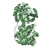
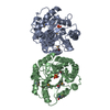
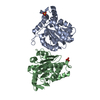
| Deposited unit | 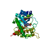
| ||||||||
|---|---|---|---|---|---|---|---|---|---|
| 1 | 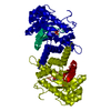
| ||||||||
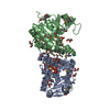
| Unit cell |
| ||||||||
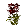
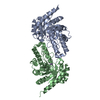
| Details | The biological assembly is a dimer constructed from chain A a symmetry partner generated by the two-fold. |
-Components
| #1: Protein | / DHPS 1 Mass: 28872.916 Da / Num. of mol.: 1 Source method: isolated from a genetically manipulated source Source: (gene. exp.) Mycobacterium tuberculosis H37Rv (bacteria)Species: Mycobacterium tuberculosis / Strain: H37RV / Plasmid: PKOS007-90 / Production host: Escherichia coli (E. coli)References: UniProt: P0A578, UniProt: P9WND1*PLUS, dihydropteroate synthase |
|---|---|
| #2: Chemical | ChemComp-MG /   Mass: 24.305 Da / Num. of mol.: 1 / Source method: obtained synthetically / Formula: Mg Mass: 24.305 Da / Num. of mol.: 1 / Source method: obtained synthetically / Formula: MgDetails: PtP was synthesized from 6-hydroxymethyl pterin and pyrophosphoric acid as described by Shiota et al., 1964 |
| #3: Chemical | ChemComp-PMM / 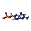  Mass: 273.143 Da / Num. of mol.: 1 / Source method: obtained synthetically / Formula: C7H8N5O5P Mass: 273.143 Da / Num. of mol.: 1 / Source method: obtained synthetically / Formula: C7H8N5O5P |
| #4: Water | ChemComp-HOH / Water Mass: 18.015 Da / Num. of mol.: 244 / Source method: isolated from a natural source / Formula: H2O Mass: 18.015 Da / Num. of mol.: 244 / Source method: isolated from a natural source / Formula: H2O |
-Experimental details
-Experiment
| Experiment | Method: X-RAY DIFFRACTION / Number of used crystals: 1 |
|---|
- Sample preparation
| Crystal | Density Matthews: 2.4 Å3/Da / Density % sol: 48.74 % | ||||||||||||||||||||||||||||||
|---|---|---|---|---|---|---|---|---|---|---|---|---|---|---|---|---|---|---|---|---|---|---|---|---|---|---|---|---|---|---|---|
| Crystal grow | Temperature: 298 K / Method: vapor diffusion, sitting drop / pH: 5.8 Details: PEG 4000, Sodium Acetate, Ammonium Acetate, para-aminosalicylic acid, 6-hydroxymethl pterin monophosphate, pH 5.8, VAPOR DIFFUSION, SITTING DROP, temperature 298K | ||||||||||||||||||||||||||||||
| Crystal grow | *PLUS Temperature: 25 ℃ | ||||||||||||||||||||||||||||||
| Components of the solutions | *PLUS
|
-Data collection
| Diffraction | Mean temperature: 140 K |
|---|---|
| Diffraction source | Source: ROTATING ANODE / Type: RIGAKU RU200 / Wavelength: 1.5418 |
| Detector | Type: RIGAKU RAXIS IIC / Detector: IMAGE PLATE / Date: Jul 4, 1999 |
| Radiation | Protocol: SINGLE WAVELENGTH / Monochromatic (M) / Laue (L): M / Scattering type: x-ray |
| Radiation wavelength | Wavelength: 1.5418 Å / Relative weight: 1 |
| Reflection | Resolution: 1.7→26.5 Å / Num. all: 136341 / Num. obs: 25211 / % possible obs: 80.5 % / Observed criterion σ(F): 2 / Observed criterion σ(I): 2 / Redundancy: 5.4 % / Biso Wilson estimate: 20.649 Å2 / Rmerge(I) obs: 0.043 / Net I/σ(I): 32.411 |
| Reflection shell | Resolution: 1.7→1.76 Å / Redundancy: 4.1 % / Rmerge(I) obs: 0.177 / Num. unique all: 2137 / % possible all: 69.9 |
| Reflection | *PLUS Num. measured all: 136341 |
| Reflection shell | *PLUS % possible obs: 69.9 % |
- Processing
| Software |
| |||||||||||||||||||||||||||||||||
|---|---|---|---|---|---|---|---|---|---|---|---|---|---|---|---|---|---|---|---|---|---|---|---|---|---|---|---|---|---|---|---|---|---|---|
| Refinement | Resolution: 1.7→20 Å / σ(F): 0 / σ(I): 0 / Stereochemistry target values: TNT default
| |||||||||||||||||||||||||||||||||
| Refinement step | Cycle: LAST / Resolution: 1.7→20 Å
| |||||||||||||||||||||||||||||||||
| Software | *PLUS Name: TNT / Classification: refinement | |||||||||||||||||||||||||||||||||
| Refinement | *PLUS % reflection Rfree: 5 % | |||||||||||||||||||||||||||||||||
| Solvent computation | *PLUS | |||||||||||||||||||||||||||||||||
| Displacement parameters | *PLUS | |||||||||||||||||||||||||||||||||
| Refine LS restraints | *PLUS
|
