 Movie
Movie Controller


+ Open data

- Basic information
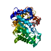
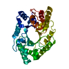
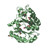
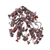
| Entry | Database: PDB / ID: 1exp | |||||||||
|---|---|---|---|---|---|---|---|---|---|---|
| Title | BETA-1,4-GLYCANASE CEX-CD | |||||||||
 Components Components | BETA-1,4-D-GLYCANASE CEX-CD | |||||||||
 Keywords Keywords |  HYDROLASE / CELLULOSE DEGRADATION / GLYCOSIDASE HYDROLASE / CELLULOSE DEGRADATION / GLYCOSIDASE | |||||||||
| Function / homology |  Function and homology information Function and homology informationcellulose 1,4-beta-cellobiosidase (non-reducing end) / cellulose 1,4-beta-cellobiosidase activity / polysaccharide binding / endo-1,4-beta-xylanase activity / endo-1,4-beta-xylanase / xylan catabolic process / cellulose catabolic processSimilarity search - Function | |||||||||
| Biological species |  Cellulomonas fimi (bacteria) Cellulomonas fimi (bacteria) | |||||||||
| Method | X-RAY DIFFRACTION / Resolution: 1.8 Å | |||||||||
 Authors Authors | White, A. / Tull, D. / Johns, K.L. / Withers, S.G. / Rose, D.R. | |||||||||
 Citation Citation | Journal: Nat.Struct.Biol. / Year: 1996 Title: Crystallographic observation of a covalent catalytic intermediate in a beta-glycosidase. Authors: White, A. / Tull, D. / Johns, K. / Withers, S.G. / Rose, D.R. #1: Journal: Biochemistry / Year: 1994Title: Crystal Structure of the Catalytic Domain of the Beta-1,4-Glycanase Cex from Cellulomonas Fimi Authors: White, A. / Withers, S.G. / Gilkes, N.R. / Rose, D.R. #2: Journal: J.Mol.Biol. / Year: 1992Title: Crystallization and Preliminary X-Ray Diffraction Analysis of the Catalytic Domain of Cex, an Exo-Beta-1,4-Glucanase and Beta-1,4-Xylanase from the Bacterium Cellulomonas Fimi Authors: Bedarkar, S. / Gilkes, N.R. / Kilburn, D.G. / Kwan, E. / Rose, D.R. / Miller Junior, R.C. / Warren, R.A. / Withers, S.G. | |||||||||
| History |
|
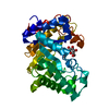
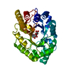
- Structure visualization
| Structure viewer | Molecule: MolmilJmol/JSmol |
|---|
- Downloads & links
-Download
| PDBx/mmCIF format | 1exp.cif.gz | 73.5 KB | Display | PDBx/mmCIF format |
|---|---|---|---|---|
| PDB format | pdb1exp.ent.gz | 57.2 KB | Display | PDB format |
| PDBx/mmJSON format | 1exp.json.gz | Tree view | PDBx/mmJSON format | |
| Others |  Other downloads Other downloads |
-Validation report
| Arichive directory | https://data.pdbj.org/pub/pdb/validation_reports/ex/1expftp://data.pdbj.org/pub/pdb/validation_reports/ex/1exp | HTTPS FTP |
|---|
-Related structure data
| Similar structure data |
|---|


-Links
 PDBj
PDBj

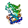
- Assembly
| Deposited unit | 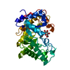
| ||||||||
|---|---|---|---|---|---|---|---|---|---|
| 1 |
| ||||||||
| Unit cell |
| ||||||||
| Components on special symmetry positions |
|
-Components
| #1: Protein | Mass: 34051.941 Da / Num. of mol.: 1 / Fragment: CATALYTICALLY ACTIVE DOMAIN Source method: isolated from a genetically manipulated source Source: (gene. exp.) Cellulomonas fimi (bacteria) / Production host: Escherichia coli (E. coli)References: GenBank: 144429, UniProt: P07986*PLUS, cellulose 1,4-beta-cellobiosidase (non-reducing end), endo-1,4-beta-xylanase |
|---|---|
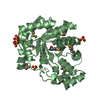
| #2: Polysaccharide | beta-D-glucopyranose-(1-4)-2-deoxy-2-fluoro-alpha-D-glucopyranose / 2-deoxy-2-fluoro-beta-cellobiose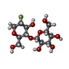  , Oligosaccharide / Class: Substrate analog / Mass: 344.288 Da / Num. of mol.: 1 , Oligosaccharide / Class: Substrate analog / Mass: 344.288 Da / Num. of mol.: 1Source method: isolated from a genetically manipulated source Details: oligosaccharide / References: 2-deoxy-2-fluoro-beta-cellobiose |
| #3: Water | ChemComp-HOH / Water Mass: 18.015 Da / Num. of mol.: 189 / Source method: isolated from a natural source / Formula: H2O Mass: 18.015 Da / Num. of mol.: 189 / Source method: isolated from a natural source / Formula: H2O |
-Experimental details
-Experiment
| Experiment | Method: X-RAY DIFFRACTION |
|---|
- Sample preparation
| Crystal | Density Matthews: 2.31 Å3/Da / Density % sol: 46.84 % | ||||||||||||||||||||||||||||||||||||
|---|---|---|---|---|---|---|---|---|---|---|---|---|---|---|---|---|---|---|---|---|---|---|---|---|---|---|---|---|---|---|---|---|---|---|---|---|---|
| Crystal grow | *PLUS pH: 4.6 / Method: vapor diffusion, hanging drop / Details: Bedarkar, S., (1992) J.Mol. Biol, 228, 693. | ||||||||||||||||||||||||||||||||||||
| Components of the solutions | *PLUS
|
-Data collection
| Diffraction source | Wavelength: 1.5418 |
|---|---|
| Detector | Type: XUONG-HAMLIN MULTIWIRE / Detector: AREA DETECTOR |
| Radiation | Monochromatic (M) / Laue (L): M / Scattering type: x-ray |
| Radiation wavelength | Wavelength: 1.5418 Å / Relative weight: 1 |
| Reflection | Resolution: 1.8→15 Å / Num. obs: 29109 / % possible obs: 99 % / Observed criterion σ(I): 0 / Redundancy: 4.7 % / Rmerge(I) obs: 0.041 |
| Reflection | *PLUS Lowest resolution: 8 Å / Num. obs: 29464 / Num. measured all: 139457 |
- Processing
| Software |
| ||||||||||||||||||||||||||||||||||||||||||||||||||||||||||||
|---|---|---|---|---|---|---|---|---|---|---|---|---|---|---|---|---|---|---|---|---|---|---|---|---|---|---|---|---|---|---|---|---|---|---|---|---|---|---|---|---|---|---|---|---|---|---|---|---|---|---|---|---|---|---|---|---|---|---|---|---|---|
| Refinement | Resolution: 1.8→8 Å / σ(F): 2
| ||||||||||||||||||||||||||||||||||||||||||||||||||||||||||||
| Refine analyze | Luzzati coordinate error obs: 0.25 Å | ||||||||||||||||||||||||||||||||||||||||||||||||||||||||||||
| Refinement step | Cycle: LAST / Resolution: 1.8→8 Å
| ||||||||||||||||||||||||||||||||||||||||||||||||||||||||||||
| Refine LS restraints |
| ||||||||||||||||||||||||||||||||||||||||||||||||||||||||||||
| Software | *PLUS Name: X-PLOR / Version: 3.1 / Classification: refinement | ||||||||||||||||||||||||||||||||||||||||||||||||||||||||||||
| Refinement | *PLUS Num. reflection obs: 24597 | ||||||||||||||||||||||||||||||||||||||||||||||||||||||||||||
| Solvent computation | *PLUS | ||||||||||||||||||||||||||||||||||||||||||||||||||||||||||||
| Displacement parameters | *PLUS | ||||||||||||||||||||||||||||||||||||||||||||||||||||||||||||
| Refine LS restraints | *PLUS
|
