 Movie
Movie Controller


+ Open data

- Basic information
| Entry | Database: PDB / ID: 1esk | ||||||
|---|---|---|---|---|---|---|---|
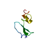
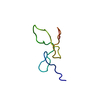
| Title | SOLUTION STRUCTURE OF NCP7 FROM HIV-1 | ||||||
 Components Components | GAG POLYPROTEIN Group-specific antigen Group-specific antigen | ||||||
 Keywords Keywords | VIRAL PROTEIN / (12-53)NCp7 / HIV-1 / PROTEIN | ||||||
| Function / homology |  Function and homology informationintegrase activity / Integration of viral DNA into host genomic DNA / Autointegration results in viral DNA circles / Minus-strand DNA synthesis / Plus-strand DNA synthesis / Uncoating of the HIV Virion / 2-LTR circle formation / Vpr-mediated nuclear import of PICs / Early Phase of HIV Life Cycle / Integration of provirus ...integrase activity / Integration of viral DNA into host genomic DNA / Autointegration results in viral DNA circles / Minus-strand DNA synthesis / Plus-strand DNA synthesis / Uncoating of the HIV Virion / 2-LTR circle formation / Vpr-mediated nuclear import of PICs / Early Phase of HIV Life Cycle / Integration of provirus / APOBEC3G mediated resistance to HIV-1 infection / Binding and entry of HIV virion / viral life cycle / Assembly Of The HIV Virion / HIV-1 retropepsin / : / Budding and maturation of HIV virion / retroviral ribonuclease H / exoribonuclease H / : / exoribonuclease H activity / protein processing / host multivesicular body / RNA-directed DNA polymerase / viral genome integration into host DNA / viral penetration into host nucleus / establishment of integrated proviral latency / RNA-directed DNA polymerase activity / Transferases; Transferring phosphorus-containing groups; Nucleotidyltransferases / RNA-DNA hybrid ribonuclease activity / peptidase activity / viral nucleocapsid / DNA recombination / Hydrolases; Acting on ester bonds / DNA-directed DNA polymerase / aspartic-type endopeptidase activity / DNA-directed DNA polymerase activity / symbiont entry into host cell / symbiont-mediated suppression of host gene expression / lipid binding / host cell nucleus / structural molecule activity / host cell plasma membrane / virion membrane / DNA binding / RNA binding / zinc ion binding / membrane / identical protein binding Function and homology informationintegrase activity / Integration of viral DNA into host genomic DNA / Autointegration results in viral DNA circles / Minus-strand DNA synthesis / Plus-strand DNA synthesis / Uncoating of the HIV Virion / 2-LTR circle formation / Vpr-mediated nuclear import of PICs / Early Phase of HIV Life Cycle / Integration of provirus ...integrase activity / Integration of viral DNA into host genomic DNA / Autointegration results in viral DNA circles / Minus-strand DNA synthesis / Plus-strand DNA synthesis / Uncoating of the HIV Virion / 2-LTR circle formation / Vpr-mediated nuclear import of PICs / Early Phase of HIV Life Cycle / Integration of provirus / APOBEC3G mediated resistance to HIV-1 infection / Binding and entry of HIV virion / viral life cycle / Assembly Of The HIV Virion / HIV-1 retropepsin / : / Budding and maturation of HIV virion / retroviral ribonuclease H / exoribonuclease H / : / exoribonuclease H activity / protein processing / host multivesicular body / RNA-directed DNA polymerase / viral genome integration into host DNA / viral penetration into host nucleus / establishment of integrated proviral latency / RNA-directed DNA polymerase activity / Transferases; Transferring phosphorus-containing groups; Nucleotidyltransferases / RNA-DNA hybrid ribonuclease activity / peptidase activity / viral nucleocapsid / DNA recombination / Hydrolases; Acting on ester bonds / DNA-directed DNA polymerase / aspartic-type endopeptidase activity / DNA-directed DNA polymerase activity / symbiont entry into host cell / symbiont-mediated suppression of host gene expression / lipid binding / host cell nucleus / structural molecule activity / host cell plasma membrane / virion membrane / DNA binding / RNA binding / zinc ion binding / membrane / identical protein bindingSimilarity search - Function | ||||||
| Method | SOLUTION NMR / simulated annealing | ||||||
 Authors Authors | Morellet, N. / Demene, H. / Teilleux, V. / Huynh-Dinh, T. / de Rocquigny, H. / Fournie-Zaluski, M.-C. / Roques, B.P. | ||||||
 Citation Citation | Journal: To be Published Title: Solution Structure of (12-53)NCp7 of HIV-1 Authors: Morellet, N. / Demene, H. / Teilleux, V. / Huynh-Dinh, T. / de Rocquigny, H. / Fournie-Zaluski, M.-C. / Roques, B.P. #1: Journal: J.Mol.Biol. / Year: 1994Title: Conformational Behaviour of the Active and Inactive Forms of the Nucleocapsid NCp7 of HIV-1 Studied by 1H NMR. Authors: Morellet, N. / de Roquigny, H. / Mely, Y. / Jullian, N. / Demene, H. / Ottmann, M. / Gerard, D. / Darlix, J.L. / Fournie-Zaluski, M.C. / Roques, B.P. #2: Journal: J.Mol.Biol. / Year: 1998Title: Structure of the Complex Between the HIV-1 Nucleocapsid Protein and the Single-stranded Pentanucleotide d(ACGCC). Authors: Morellet, N. / Demene, H. / Teilleux, V. / Huynh-Dinh, T. / de Roquigny, H. / Fournie-Zaluski, M.C. / Roques, B.P. | ||||||
| History |
|
- Structure visualization
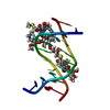
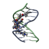
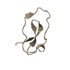
| Structure viewer | Molecule: MolmilJmol/JSmol |
|---|
- Downloads & links
-Download
| PDBx/mmCIF format | 1esk.cif.gz | 122 KB | Display | PDBx/mmCIF format |
|---|---|---|---|---|
| PDB format | pdb1esk.ent.gz | 104 KB | Display | PDB format |
| PDBx/mmJSON format | 1esk.json.gz | Tree view | PDBx/mmJSON format | |
| Others |  Other downloads Other downloads |
-Validation report
| Arichive directory | https://data.pdbj.org/pub/pdb/validation_reports/es/1eskftp://data.pdbj.org/pub/pdb/validation_reports/es/1esk | HTTPS FTP |
|---|
-Related structure data
| Related structure data | |
|---|---|
| Similar structure data |


-Links
 PDBj
PDBj


- Assembly
| Deposited unit | 
| |||||||||
|---|---|---|---|---|---|---|---|---|---|---|
| 1 |
| |||||||||
| NMR ensembles |
|
-Components
| #1: Protein/peptide | Group-specific antigen / NUCLEOCAPSID PROTEIN NCP7 Mass: 4837.642 Da / Num. of mol.: 1 / Fragment: RESIDUES 12-53 / Source method: obtained synthetically Details: The protein was chemically synthesized. The sequence is naturally found in human immunodeficiency virus type 1 (HIV-1). References: UniProt: P04585 |
|---|---|
| #2: Chemical |   Mass: 65.409 Da / Num. of mol.: 2 / Source method: obtained synthetically / Formula: Zn Mass: 65.409 Da / Num. of mol.: 2 / Source method: obtained synthetically / Formula: Zn |
-Experimental details
-Experiment
| Experiment | Method: SOLUTION NMR | ||||||||||||||||
|---|---|---|---|---|---|---|---|---|---|---|---|---|---|---|---|---|---|
| NMR experiment |
| ||||||||||||||||
| NMR details | Text: This structure was determined using standard 2D homonuclear techniques. |
- Sample preparation
| Details | Contents: 2mM (12-53)NCp7, 90%H2O, 10% D2O, pH 6.0 / Solvent system: 90% H2O/10% D2O |
|---|---|
| Sample conditions | Ionic strength: n.a. / pH: 6.0 / Pressure: ambient / Temperature: 293 K |
-NMR measurement
| NMR spectrometer | Type: Bruker AMX / Manufacturer: Bruker / Model: AMX / Field strength: 600 MHz |
|---|
- Processing
| NMR software |
| ||||||||||||
|---|---|---|---|---|---|---|---|---|---|---|---|---|---|
| Refinement | Method: simulated annealing / Software ordinal: 1 Details: the structures are based on a total of 444 NOE-derived distance constraints | ||||||||||||
| NMR representative | Selection criteria: lowest energy | ||||||||||||
| NMR ensemble | Conformer selection criteria: structures with acceptable covalent geometry,structures with favorable non-bond energy,structures with the least restraint violations,structures with the lowest energy Conformers calculated total number: 50 / Conformers submitted total number: 9 |
