 Movie
Movie Controller


+ Open data

- Basic information
| Entry | Database: PDB / ID: 1ehw | ||||||
|---|---|---|---|---|---|---|---|
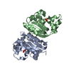
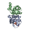
| Title | HUMAN NUCLEOSIDE DIPHOSPHATE KINASE 4 | ||||||
 Components Components | NUCLEOSIDE DIPHOSPHATE KINASE Nucleoside-diphosphate kinase Nucleoside-diphosphate kinase | ||||||
 Keywords Keywords | TRANSFERASE / nucleoside diphosphate kinase / nm23 / mitochondrial / killer-of-prune | ||||||
| Function / homology |  Function and homology information Function and homology informationnucleoside metabolic process / cardiolipin binding / nucleobase-containing small molecule interconversion / nucleoside-diphosphate kinase / Interconversion of nucleotide di- and triphosphates / UTP biosynthetic process / CTP biosynthetic process / GTP biosynthetic process / lipid transport / nucleoside diphosphate kinase activity ...nucleoside metabolic process / cardiolipin binding / nucleobase-containing small molecule interconversion / nucleoside-diphosphate kinase / Interconversion of nucleotide di- and triphosphates / UTP biosynthetic process / CTP biosynthetic process / GTP biosynthetic process / lipid transport / nucleoside diphosphate kinase activity / mitochondrial intermembrane space / mitochondrial inner membrane / mitochondrial matrix / mitochondrion / ATP binding / metal ion bindingSimilarity search - Function | ||||||
| Biological species |  Homo sapiens (human) Homo sapiens (human) | ||||||
| Method | X-RAY DIFFRACTION / SYNCHROTRON / Resolution: 2.4 Å | ||||||
 Authors Authors | Milon, L. / Meyer, P. / Chiadmi, M. / Munier, A. / Johansson, M. / Karlsson, A. / Lascu, I. / Capeau, J. / Janin, J. / Lacombe, M.-L. | ||||||
 Citation Citation | Journal: J.Biol.Chem. / Year: 2000 Title: The human nm23-H4 gene product is a mitochondrial nucleoside diphosphate kinase. Authors: Milon, L. / Meyer, P. / Chiadmi, M. / Munier, A. / Johansson, M. / Karlsson, A. / Lascu, I. / Capeau, J. / Janin, J. / Lacombe, M.L. | ||||||
| History |
|
- Structure visualization
| Structure viewer | Molecule: MolmilJmol/JSmol |
|---|
- Downloads & links
-Download
| PDBx/mmCIF format | 1ehw.cif.gz | 70.5 KB | Display | PDBx/mmCIF format |
|---|---|---|---|---|
| PDB format | pdb1ehw.ent.gz | 52.8 KB | Display | PDB format |
| PDBx/mmJSON format | 1ehw.json.gz | Tree view | PDBx/mmJSON format | |
| Others |  Other downloads Other downloads |
-Validation report
| Arichive directory | https://data.pdbj.org/pub/pdb/validation_reports/eh/1ehwftp://data.pdbj.org/pub/pdb/validation_reports/eh/1ehw | HTTPS FTP |
|---|
-Related structure data
| Similar structure data |
|---|


-Links
 PDBj
PDBj

- Assembly
| Deposited unit | 
| ||||||||
|---|---|---|---|---|---|---|---|---|---|
| 1 | 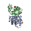
| ||||||||
| Unit cell |
| ||||||||
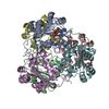
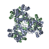
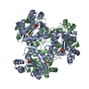
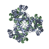
| Details | The biological assembly is an hexamer constructed from the dimer formed by chain A and B generated by the three-fold |
-Components
| #1: Protein | Nucleoside-diphosphate kinase / NDPK H4 Mass: 18209.717 Da / Num. of mol.: 2 / Mutation: S34M, W35G Source method: isolated from a genetically manipulated source Details: N-TERMINUS TRUNCATED, N-TERMINAL HIS TAG / Source: (gene. exp.) Homo sapiens (human) / Plasmid: PLASMID PET-28A(+) NOVAGEN / Production host:  Escherichia coli (E. coli) / References: UniProt: O00746, nucleoside-diphosphate kinase Escherichia coli (E. coli) / References: UniProt: O00746, nucleoside-diphosphate kinase#2: Chemical | Sulfate  Mass: 96.063 Da / Num. of mol.: 3 / Source method: obtained synthetically / Formula: SO4 Mass: 96.063 Da / Num. of mol.: 3 / Source method: obtained synthetically / Formula: SO4#3: Water | ChemComp-HOH / | Water Mass: 18.015 Da / Num. of mol.: 83 / Source method: isolated from a natural source / Formula: H2O Mass: 18.015 Da / Num. of mol.: 83 / Source method: isolated from a natural source / Formula: H2O |
|---|
-Experimental details
-Experiment
| Experiment | Method: X-RAY DIFFRACTION / Number of used crystals: 1 |
|---|
- Sample preparation
| Crystal | Density Matthews: 3.3 Å3/Da / Density % sol: 62.69 % | ||||||||||||||||||||||||||||||||||||||||||||||||||||||||||||
|---|---|---|---|---|---|---|---|---|---|---|---|---|---|---|---|---|---|---|---|---|---|---|---|---|---|---|---|---|---|---|---|---|---|---|---|---|---|---|---|---|---|---|---|---|---|---|---|---|---|---|---|---|---|---|---|---|---|---|---|---|---|
| Crystal grow | Temperature: 291 K / Method: vapor diffusion, hanging drop / pH: 7.5 Details: lithium sulfate, sodium chloride, HEPES, EDTA, pH 7.5, VAPOR DIFFUSION, HANGING DROP, temperature 291K | ||||||||||||||||||||||||||||||||||||||||||||||||||||||||||||
| Crystal grow | *PLUS | ||||||||||||||||||||||||||||||||||||||||||||||||||||||||||||
| Components of the solutions | *PLUS
|
-Data collection
| Diffraction | Mean temperature: 277 K |
|---|---|
| Diffraction source | Source: SYNCHROTRON / Site: LURE  / Beamline: DW32 / Wavelength: 0.965 / Beamline: DW32 / Wavelength: 0.965 |
| Detector | Type: MARRESEARCH / Detector: IMAGE PLATE |
| Radiation | Protocol: SINGLE WAVELENGTH / Monochromatic (M) / Laue (L): M / Scattering type: x-ray |
| Radiation wavelength | Wavelength: 0.965 Å / Relative weight: 1 |
| Reflection | Resolution: 2.4→30 Å / Num. obs: 19054 / Biso Wilson estimate: 29.5 Å2 |
| Reflection | *PLUS % possible obs: 94.3 % / Num. measured all: 115939 / Rmerge(I) obs: 0.105 |
- Processing
| Software |
| ||||||||||||||||||||||||||||||||||||||||||||||||||||||||||||||||||||||||||||||||
|---|---|---|---|---|---|---|---|---|---|---|---|---|---|---|---|---|---|---|---|---|---|---|---|---|---|---|---|---|---|---|---|---|---|---|---|---|---|---|---|---|---|---|---|---|---|---|---|---|---|---|---|---|---|---|---|---|---|---|---|---|---|---|---|---|---|---|---|---|---|---|---|---|---|---|---|---|---|---|---|---|---|
| Refinement | Resolution: 2.4→30 Å / Rfactor Rfree error: 0.008 / Data cutoff high absF: 3814914.74 / Data cutoff low absF: 0 / Isotropic thermal model: RESTRAINED / Cross valid method: THROUGHOUT / σ(F): 0
| ||||||||||||||||||||||||||||||||||||||||||||||||||||||||||||||||||||||||||||||||
| Solvent computation | Solvent model: FLAT MODEL / Bsol: 40.55 Å2 / ksol: 0.351 e/Å3 | ||||||||||||||||||||||||||||||||||||||||||||||||||||||||||||||||||||||||||||||||
| Displacement parameters | Biso mean: 34.5 Å2
| ||||||||||||||||||||||||||||||||||||||||||||||||||||||||||||||||||||||||||||||||
| Refine analyze |
| ||||||||||||||||||||||||||||||||||||||||||||||||||||||||||||||||||||||||||||||||
| Refinement step | Cycle: LAST / Resolution: 2.4→30 Å
| ||||||||||||||||||||||||||||||||||||||||||||||||||||||||||||||||||||||||||||||||
| Refine LS restraints |
| ||||||||||||||||||||||||||||||||||||||||||||||||||||||||||||||||||||||||||||||||
| LS refinement shell | Resolution: 2.4→2.55 Å / Rfactor Rfree error: 0.026 / Total num. of bins used: 6
| ||||||||||||||||||||||||||||||||||||||||||||||||||||||||||||||||||||||||||||||||
| Xplor file |
| ||||||||||||||||||||||||||||||||||||||||||||||||||||||||||||||||||||||||||||||||
| Software | *PLUS Name: CNS / Version: 0.5 / Classification: refinement | ||||||||||||||||||||||||||||||||||||||||||||||||||||||||||||||||||||||||||||||||
| Refinement | *PLUS σ(F): 0 / % reflection Rfree: 5.1 % | ||||||||||||||||||||||||||||||||||||||||||||||||||||||||||||||||||||||||||||||||
| Solvent computation | *PLUS | ||||||||||||||||||||||||||||||||||||||||||||||||||||||||||||||||||||||||||||||||
| Displacement parameters | *PLUS Biso mean: 34.5 Å2 | ||||||||||||||||||||||||||||||||||||||||||||||||||||||||||||||||||||||||||||||||
| Refine LS restraints | *PLUS
| ||||||||||||||||||||||||||||||||||||||||||||||||||||||||||||||||||||||||||||||||
| LS refinement shell | *PLUS Rfactor Rfree: 0.323 / % reflection Rfree: 5 % / Rfactor Rwork: 0.244 |
