 Movie
Movie Controller


+ Open data

- Basic information
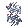
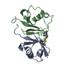
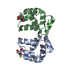
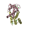
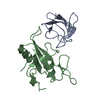
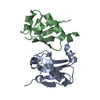
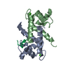
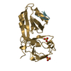
| Entry | Database: PDB / ID: 1e8a | ||||||
|---|---|---|---|---|---|---|---|
| Title | The three-dimensional structure of human S100A12 | ||||||
 Components Components | S100A12 | ||||||
 Keywords Keywords | ANTIFUNGAL PROTEIN / S100 PROTEIN / EF-HAND / CALCIUM BINDING | ||||||
| Function / homology |  Function and homology information Function and homology informationmast cell activation / RAGE receptor binding / monocyte chemotaxis / TRAF6 mediated NF-kB activation / Advanced glycosylation endproduct receptor signaling / endothelial cell migration / defense response to fungus / xenobiotic metabolic process / neutrophil chemotaxis / TAK1-dependent IKK and NF-kappa-B activation ...mast cell activation / RAGE receptor binding / monocyte chemotaxis / TRAF6 mediated NF-kB activation / Advanced glycosylation endproduct receptor signaling / endothelial cell migration / defense response to fungus / xenobiotic metabolic process / neutrophil chemotaxis / TAK1-dependent IKK and NF-kappa-B activation / positive regulation of MAP kinase activity / positive regulation of inflammatory response / calcium-dependent protein binding / antimicrobial humoral immune response mediated by antimicrobial peptide / positive regulation of NF-kappaB transcription factor activity / positive regulation of canonical NF-kappaB signal transduction / secretory granule lumen / killing of cells of another organism / cytoskeleton / defense response to bacterium / inflammatory response / copper ion binding / innate immune response / calcium ion binding / Neutrophil degranulation / zinc ion binding / extracellular region / identical protein binding / nucleus / plasma membrane / cytosol / cytoplasmSimilarity search - Function | ||||||
| Biological species |  HOMO SAPIENS (human) HOMO SAPIENS (human) | ||||||
| Method | X-RAY DIFFRACTION / SYNCHROTRON / MOLECULAR REPLACEMENT / Resolution: 1.95 Å | ||||||
 Authors Authors | Moroz, O.V. / Antson, A.A. / Murshudov, G.N. / Maitland, N.J. / Dodson, G.G. / Wilson, K.S. / Skibshoj, I. / Lukanidin, E.M. / Bronstein, I.B. | ||||||
 Citation Citation | Journal: Acta Crystallogr.,Sect.D / Year: 2001 Title: The Three-Dimensional Structure of Human S100A12 Authors: Moroz, O.V. / Antson, A.A. / Murshudov, G.N. / Maitland, N.J. / Dodson, G.G. / Wilson, K.S. / Skibshoj, I. / Lukanidin, E.M. / Bronstein, I.B. #1: Journal: Acta Crystallogr.,Sect.D / Year: 2000 Title: Crystallization and Preliminary X-Ray Diffraction Analysis of Human Calcium-Binding Protein S100A12 Authors: Moroz, O.V. / Antson, A.A. / Dodson, G.G. / Wilson, K.S. / Skibshoj, I. / Lukanidin, E. / Bronstein, I. | ||||||
| History |
|
- Structure visualization
| Structure viewer | Molecule: MolmilJmol/JSmol |
|---|
- Downloads & links
-Download
| PDBx/mmCIF format | 1e8a.cif.gz | 53 KB | Display | PDBx/mmCIF format |
|---|---|---|---|---|
| PDB format | pdb1e8a.ent.gz | 37.5 KB | Display | PDB format |
| PDBx/mmJSON format | 1e8a.json.gz | Tree view | PDBx/mmJSON format | |
| Others |  Other downloads Other downloads |
-Validation report
| Arichive directory | https://data.pdbj.org/pub/pdb/validation_reports/e8/1e8aftp://data.pdbj.org/pub/pdb/validation_reports/e8/1e8a | HTTPS FTP |
|---|
-Related structure data
| Related structure data |  1mhoS S: Starting model for refinement |
|---|---|
| Similar structure data |


-Links
 PDBj
PDBj


- Assembly
| Deposited unit | 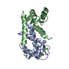
| ||||||||
|---|---|---|---|---|---|---|---|---|---|
| 1 |
| ||||||||
| Unit cell |
|
-Components
| #1: Protein | / CALGRANULIN C Mass: 10460.834 Da / Num. of mol.: 2 / Source method: isolated from a natural source / Source: (natural) HOMO SAPIENS (human) / Cell: GRANULOCYTE / Tissue: BLOOD / References: UniProt: P80511#2: Chemical | ChemComp-CA /   Mass: 40.078 Da / Num. of mol.: 4 / Source method: obtained synthetically / Formula: Ca Mass: 40.078 Da / Num. of mol.: 4 / Source method: obtained synthetically / Formula: Ca#3: Water | ChemComp-HOH / | Water Mass: 18.015 Da / Num. of mol.: 180 / Source method: isolated from a natural source / Formula: H2O Mass: 18.015 Da / Num. of mol.: 180 / Source method: isolated from a natural source / Formula: H2O |
|---|
-Experimental details
-Experiment
| Experiment | Method: X-RAY DIFFRACTION / Number of used crystals: 1 |
|---|
- Sample preparation
| Crystal | Density Matthews: 2.9 Å3/Da / Density % sol: 57.9 % | |||||||||||||||||||||||||||||||||||
|---|---|---|---|---|---|---|---|---|---|---|---|---|---|---|---|---|---|---|---|---|---|---|---|---|---|---|---|---|---|---|---|---|---|---|---|---|
| Crystal grow | Method: vapor diffusion, hanging drop / pH: 6.5 Details: 20-25% PEG 5K MONOMETHYL ETHER, 0.2M CALCIUM CHLORIDE, 0.1M SODIUM CACODILATE, HANGING DROPS, pH 6.50 | |||||||||||||||||||||||||||||||||||
| Crystal grow | *PLUS Method: vapor diffusion, hanging drop / pH: 6.5 | |||||||||||||||||||||||||||||||||||
| Components of the solutions | *PLUS
|
-Data collection
| Diffraction | Mean temperature: 120 K |
|---|---|
| Diffraction source | Source: SYNCHROTRON / Site: ESRF  / Beamline: ID14-4 / Wavelength: 0.93 / Beamline: ID14-4 / Wavelength: 0.93 |
| Radiation | Protocol: SINGLE WAVELENGTH / Monochromatic (M) / Laue (L): M / Scattering type: x-ray |
| Radiation wavelength | Wavelength: 0.93 Å / Relative weight: 1 |
| Reflection | Resolution: 1.95→25 Å / Num. obs: 17174 / % possible obs: 99.3 % / Redundancy: 2.8 % / Rmerge(I) obs: 0.086 / Net I/σ(I): 10.6 |
| Reflection shell | Resolution: 1.95→2.02 Å / Redundancy: 1.8 % / Rmerge(I) obs: 0.25 / Mean I/σ(I) obs: 2.68 / % possible all: 99.1 |
| Reflection shell | *PLUS % possible obs: 99.1 % / Num. unique obs: 1721 / Rmerge(I) obs: 0.258 |
- Processing
| Software |
| ||||||||||||||||||||||||||||||||||||||||||||||||||||||||||||||||||||||||||||||||||||
|---|---|---|---|---|---|---|---|---|---|---|---|---|---|---|---|---|---|---|---|---|---|---|---|---|---|---|---|---|---|---|---|---|---|---|---|---|---|---|---|---|---|---|---|---|---|---|---|---|---|---|---|---|---|---|---|---|---|---|---|---|---|---|---|---|---|---|---|---|---|---|---|---|---|---|---|---|---|---|---|---|---|---|---|---|---|
| Refinement | Method to determine structure: MOLECULAR REPLACEMENT Starting model: PDB ENTRY 1MHO Resolution: 1.95→25 Å / SU B: 4.36111 / SU ML: 0.12704 / Cross valid method: THROUGHOUT / σ(F): 0 / ESU R: 0.28567 / ESU R Free: 0.19523
| ||||||||||||||||||||||||||||||||||||||||||||||||||||||||||||||||||||||||||||||||||||
| Displacement parameters | Biso mean: 18.851 Å2
| ||||||||||||||||||||||||||||||||||||||||||||||||||||||||||||||||||||||||||||||||||||
| Refinement step | Cycle: LAST / Resolution: 1.95→25 Å
| ||||||||||||||||||||||||||||||||||||||||||||||||||||||||||||||||||||||||||||||||||||
| Refine LS restraints |
| ||||||||||||||||||||||||||||||||||||||||||||||||||||||||||||||||||||||||||||||||||||
| Software | *PLUS Name: REFMAC / Classification: refinement | ||||||||||||||||||||||||||||||||||||||||||||||||||||||||||||||||||||||||||||||||||||
| Refinement | *PLUS Rfactor obs: 0.178 | ||||||||||||||||||||||||||||||||||||||||||||||||||||||||||||||||||||||||||||||||||||
| Solvent computation | *PLUS | ||||||||||||||||||||||||||||||||||||||||||||||||||||||||||||||||||||||||||||||||||||
| Displacement parameters | *PLUS Biso mean: 18.851 Å2 |
