 Movie
Movie Controller

[English] 日本語
 Yorodumi
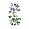
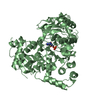
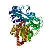
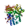
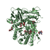
- PDB-1e6e: ADRENODOXIN REDUCTASE/ADRENODOXIN COMPLEX OF MITOCHONDRIAL P450 S... -
+ Open data

- Basic information
| Entry | Database: PDB / ID: 1e6e | ||||||
|---|---|---|---|---|---|---|---|
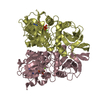
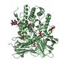
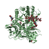
| Title | ADRENODOXIN REDUCTASE/ADRENODOXIN COMPLEX OF MITOCHONDRIAL P450 SYSTEMS | ||||||
 Components Components |
| ||||||
 Keywords Keywords |  OXIDOREDUCTASE / FLAVOENZYME / ELECTRON TRANSFERASE / [2FE-2S]FERREDOXIN / ADRENODOXIN / ELECTRON TRANSPORT / COMPLEX OXIDOREDUCTASE / FLAVOENZYME / ELECTRON TRANSFERASE / [2FE-2S]FERREDOXIN / ADRENODOXIN / ELECTRON TRANSPORT / COMPLEX | ||||||
| Function / homology |  Function and homology informationadrenodoxin-NADP+ reductase / NADPH-adrenodoxin reductase activity / Mitochondrial iron-sulfur cluster biogenesis / Pregnenolone biosynthesis / Endogenous sterols / Electron transport from NADPH to Ferredoxin / hormone biosynthetic process / P450-containing electron transport chain / ubiquinone biosynthetic process / steroid biosynthetic process ...adrenodoxin-NADP+ reductase / NADPH-adrenodoxin reductase activity / Mitochondrial iron-sulfur cluster biogenesis / Pregnenolone biosynthesis / Endogenous sterols / Electron transport from NADPH to Ferredoxin / hormone biosynthetic process / P450-containing electron transport chain / ubiquinone biosynthetic process / steroid biosynthetic process / electron transport chain / cellular response to forskolin / cellular response to cAMP / cholesterol metabolic process / respiratory electron transport chain / 2 iron, 2 sulfur cluster binding / flavin adenine dinucleotide binding / NADP binding / mitochondrial inner membrane / electron transfer activity / mitochondrial matrix / protein homodimerization activity / mitochondrion / metal ion binding Function and homology informationadrenodoxin-NADP+ reductase / NADPH-adrenodoxin reductase activity / Mitochondrial iron-sulfur cluster biogenesis / Pregnenolone biosynthesis / Endogenous sterols / Electron transport from NADPH to Ferredoxin / hormone biosynthetic process / P450-containing electron transport chain / ubiquinone biosynthetic process / steroid biosynthetic process ...adrenodoxin-NADP+ reductase / NADPH-adrenodoxin reductase activity / Mitochondrial iron-sulfur cluster biogenesis / Pregnenolone biosynthesis / Endogenous sterols / Electron transport from NADPH to Ferredoxin / hormone biosynthetic process / P450-containing electron transport chain / ubiquinone biosynthetic process / steroid biosynthetic process / electron transport chain / cellular response to forskolin / cellular response to cAMP / cholesterol metabolic process / respiratory electron transport chain / 2 iron, 2 sulfur cluster binding / flavin adenine dinucleotide binding / NADP binding / mitochondrial inner membrane / electron transfer activity / mitochondrial matrix / protein homodimerization activity / mitochondrion / metal ion bindingSimilarity search - Function | ||||||
| Biological species |  BOS TAURUS (cattle) BOS TAURUS (cattle) | ||||||
| Method | X-RAY DIFFRACTION / SYNCHROTRON / MOLECULAR REPLACEMENT / Resolution: 2.3 Å | ||||||
 Authors Authors | Mueller, J.J. / Lapko, A. / Bourenkov, G. / Ruckpaul, K. / Heinemann, U. | ||||||
 Citation Citation | Journal: J.Biol.Chem. / Year: 2001 Title: Adrenodoxin Reductase-Adrenodoxin Complex Structure Suggests Electron Transfer Path in Steroid Biosynthesis. Authors: Mueller, J.J. / Lapko, A. / Bourenkov, G. / Ruckpaul, K. / Heinemann, U. #1: Journal: To be PublishedTitle: X-Ray Structure of Bovine Adrenodoxin Reductase -Adrenodoxin Complex at 2.5 A Resolution: The First Three-Dimensional Structure of a Complex of Two Components of the Steroidogenic Electron ...Title: X-Ray Structure of Bovine Adrenodoxin Reductase -Adrenodoxin Complex at 2.5 A Resolution: The First Three-Dimensional Structure of a Complex of Two Components of the Steroidogenic Electron Transport System in Adrenal Cortex Mitochondria Authors: Mueller, J.J. / Lapko, A. / Bourenkov, G. / Mueller, E.C. / Otto, A. / Ruckpaul, K. / Heinemann, U. #2: Journal: Proteins: Struct.,Funct., Genet. / Year: 1997 Title: Preparation and Crystallization of a Cross-Linked Complex of Bovine Adrenodoxin and Adrenodoxin Reductase Authors: Lapko, A. / Mueller, A. / Heese, O. / Ruckpaul, K. / Heinemann, U. | ||||||
| History |
|
- Structure visualization
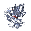
| Structure viewer | Molecule: MolmilJmol/JSmol |
|---|
- Downloads & links
-Download
| PDBx/mmCIF format | 1e6e.cif.gz | 237 KB | Display | PDBx/mmCIF format |
|---|---|---|---|---|
| PDB format | pdb1e6e.ent.gz | 189.6 KB | Display | PDB format |
| PDBx/mmJSON format | 1e6e.json.gz | Tree view | PDBx/mmJSON format | |
| Others |  Other downloads Other downloads |
-Validation report
| Arichive directory | https://data.pdbj.org/pub/pdb/validation_reports/e6/1e6eftp://data.pdbj.org/pub/pdb/validation_reports/e6/1e6e | HTTPS FTP |
|---|
-Related structure data


-Links
 PDBj
PDBj


- Assembly
| Deposited unit | 
| ||||||||
|---|---|---|---|---|---|---|---|---|---|
| 1 | 
| ||||||||
| 2 | 
| ||||||||
| Unit cell |
|
-Components
-Protein , 2 types, 4 molecules ACBD
| #1: Protein | Mass: 50360.410 Da / Num. of mol.: 2 Source method: isolated from a genetically manipulated source Details: COVALENT CROSSLINK BETWEEN LYS27 OF ADR AND ASP39 OF ADX Source: (gene. exp.) BOS TAURUS (cattle) / Tissue: STEROIDOGENIC TISSUES / Cell: MITOCHONDRION / Cellular location: ATTACHED TO INNER MITOCHONDRIAL MEMBRANE / Organ: ADRENAL GLAND / Organelle: MITOCHONDRIAL MATRIX / Plasmid: PBAR1607 / Production host:  ESCHERICHIA COLI (E. coli) / Strain (production host): BL21 ESCHERICHIA COLI (E. coli) / Strain (production host): BL21References: UniProt: P08165, ferredoxin-NADP+ reductase, adrenodoxin-NADP+ reductase#2: Protein | Adrenal ferredoxin / ADRENAL FERREDOXIN / FERREDOXIN-1 / HEPATO-FERREDOXIN / ADX / A DRENODOXINMass: 14031.734 Da / Num. of mol.: 2 / Mutation: YES Source method: isolated from a genetically manipulated source Details: COVALENT CROSSLINK BETWEEN LYS27 OF ADR AND ASP39 OF ADX Source: (gene. exp.) BOS TAURUS (cattle) / Tissue: STEROIDOGENIC TISSUES / Cell: MITOCHONDRION / Organ: ADRENAL GLAND / Organelle: MITOCHONDRIAL MATRIX / Plasmid: PKKHC, PMIXT / Production host: ESCHERICHIA COLI (E. coli) / Strain (production host): BL21 / References: UniProt: P00257 |
|---|
-Non-polymers , 4 types, 283 molecules 


| #3: Chemical | Flavin adenine dinucleotide Mass: 785.550 Da / Num. of mol.: 2 / Source method: obtained synthetically / Formula: C27H33N9O15P2 / Comment: FAD*YM Mass: 785.550 Da / Num. of mol.: 2 / Source method: obtained synthetically / Formula: C27H33N9O15P2 / Comment: FAD*YM#4: Chemical | ChemComp-SO4 / Sulfate Mass: 96.063 Da / Num. of mol.: 5 / Source method: obtained synthetically / Formula: SO4 Mass: 96.063 Da / Num. of mol.: 5 / Source method: obtained synthetically / Formula: SO4#5: Chemical | Iron–sulfur cluster Mass: 175.820 Da / Num. of mol.: 2 / Source method: obtained synthetically / Formula: Fe2S2 Mass: 175.820 Da / Num. of mol.: 2 / Source method: obtained synthetically / Formula: Fe2S2#6: Water | ChemComp-HOH / | WaterMass: 18.015 Da / Num. of mol.: 274 / Source method: isolated from a natural source / Formula: H2O |
|---|
-Details
| Compound details | CHAIN B, D ENGINEERED MUTATION SER1GLY REDUCED ADRENODOXIN + NADP(+) = OXIDIZED ADRENODOXIN + NADPH ...CHAIN B, D ENGINEERED |
|---|---|
| Sequence details | FIRST 32 RESIDUES OF ADR SWISS-PROT SEQUENCE REFER TO A MITOCHONDRIAL LEADER SEQUENCE THAT WAS ...FIRST 32 RESIDUES OF ADR SWISS-PROT SEQUENCE REFER TO A MITOCHONDR |
-Experimental details
-Experiment
| Experiment | Method: X-RAY DIFFRACTION / Number of used crystals: 2 |
|---|
- Sample preparation
| Crystal | Density Matthews: 2.9 Å3/Da / Density % sol: 57 % | ||||||||||||||||||||||||||||||
|---|---|---|---|---|---|---|---|---|---|---|---|---|---|---|---|---|---|---|---|---|---|---|---|---|---|---|---|---|---|---|---|
| Crystal grow | Temperature: 277 K / Method: vapor diffusion, hanging drop / pH: 7.4 Details: HANGING DROP VAPOR-DIFFUSION AT 4 DEGR. 0.5MM COMPLEX IN 100MMTRIS-HCL, PH 7.4, 0.9M AMMONIUMSULFATE. | ||||||||||||||||||||||||||||||
| Crystal grow | *PLUS Temperature: 4 ℃ / Method: vapor diffusion, hanging dropDetails: Lapko, A., (1997) Proteins: Struct.,Funct., Genet., 28, 289. | ||||||||||||||||||||||||||||||
| Components of the solutions | *PLUS
|
-Data collection
| Diffraction | Mean temperature: 100 K |
|---|---|
| Diffraction source | Source: SYNCHROTRON / Site: MPG/DESY, HAMBURG  / Beamline: BW6 / Wavelength: 1.046 / Beamline: BW6 / Wavelength: 1.046 |
| Detector | Type: MAR scanner 345 mm plate / Detector: IMAGE PLATE / Date: Apr 15, 1999 |
| Radiation | Protocol: SINGLE WAVELENGTH / Monochromatic (M) / Laue (L): M / Scattering type: x-ray |
| Radiation wavelength | Wavelength: 1.046 Å / Relative weight: 1 |
| Reflection | Resolution: 2.3→20 Å / Num. obs: 55229 / % possible obs: 79.1 % / Redundancy: 3.7 % / Biso Wilson estimate: 26.3 Å2 / Rsym value: 0.069 / Net I/σ(I): 18.5 |
| Reflection shell | Resolution: 2.3→2.4 Å / Mean I/σ(I) obs: 11 / Rsym value: 0.11 / % possible all: 56 |
| Reflection | *PLUS Num. measured all: 201839 / Rmerge(I) obs: 0.069 |
| Reflection shell | *PLUS % possible obs: 56 % / Rmerge(I) obs: 0.12 |
- Processing
| Software |
| ||||||||||||||||||||||||||||||||||||||||||||||||||||||||||||
|---|---|---|---|---|---|---|---|---|---|---|---|---|---|---|---|---|---|---|---|---|---|---|---|---|---|---|---|---|---|---|---|---|---|---|---|---|---|---|---|---|---|---|---|---|---|---|---|---|---|---|---|---|---|---|---|---|---|---|---|---|---|
| Refinement | Method to determine structure: MOLECULAR REPLACEMENT Starting model: PDB ENTRIES 1AYF AND 1CJC Resolution: 2.3→19.97 Å / Rfactor Rfree error: 0.005 / Data cutoff high absF: 4745426 / Isotropic thermal model: RESTRAINED / Cross valid method: THROUGHOUT / σ(F): 0
| ||||||||||||||||||||||||||||||||||||||||||||||||||||||||||||
| Solvent computation | Solvent model: FLAT MODEL / Bsol: 26.9964 Å2 / ksol: 0.348512 e/Å3 | ||||||||||||||||||||||||||||||||||||||||||||||||||||||||||||
| Displacement parameters | Biso mean: 30.4 Å2
| ||||||||||||||||||||||||||||||||||||||||||||||||||||||||||||
| Refine analyze |
| ||||||||||||||||||||||||||||||||||||||||||||||||||||||||||||
| Refinement step | Cycle: LAST / Resolution: 2.3→19.97 Å
| ||||||||||||||||||||||||||||||||||||||||||||||||||||||||||||
| Refine LS restraints |
| ||||||||||||||||||||||||||||||||||||||||||||||||||||||||||||
| LS refinement shell | Resolution: 2.3→2.44 Å / Rfactor Rfree error: 0.02 / Total num. of bins used: 6
| ||||||||||||||||||||||||||||||||||||||||||||||||||||||||||||
| Xplor file |
| ||||||||||||||||||||||||||||||||||||||||||||||||||||||||||||
| Software | *PLUS Name: CNS / Version: 1 / Classification: refinement | ||||||||||||||||||||||||||||||||||||||||||||||||||||||||||||
| Refinement | *PLUS % reflection Rfree: 5 % / Rfactor obs: 0.223 | ||||||||||||||||||||||||||||||||||||||||||||||||||||||||||||
| Solvent computation | *PLUS | ||||||||||||||||||||||||||||||||||||||||||||||||||||||||||||
| Displacement parameters | *PLUS | ||||||||||||||||||||||||||||||||||||||||||||||||||||||||||||
| Refine LS restraints | *PLUS
| ||||||||||||||||||||||||||||||||||||||||||||||||||||||||||||
| LS refinement shell | *PLUS Highest resolution: 2.3 Å |
