 Movie
Movie Controller

[English] 日本語
 Yorodumi
- PDB-1e5k: CRYSTAL STRUCTURE OF THE MOLYBDENUM COFACTOR BIOSYNTHESIS PROTEIN... -
+ Open data

- Basic information
| Entry | Database: PDB / ID: 1e5k | ||||||
|---|---|---|---|---|---|---|---|
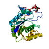
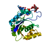
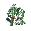
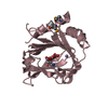
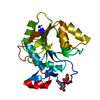
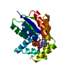
| Title | CRYSTAL STRUCTURE OF THE MOLYBDENUM COFACTOR BIOSYNTHESIS PROTEIN MOBA (PROTEIN FA) FROM ESCHERICHIA COLI AT NEAR ATOMIC RESOLUTION | ||||||
 Components Components | MOLYBDOPTERIN-GUANINE DINUCLEOTIDE BIOSYNTHESIS PROTEIN A | ||||||
 Keywords Keywords | MOLYBDOPTERIN NUCLEOTIDYL-TRANSFERASE | ||||||
| Function / homology |  Function and homology information Function and homology informationbis(molybdopterin guanine dinucleotide)molybdenum biosynthetic process /  molybdenum cofactor guanylyltransferase / molybdenum cofactor guanylyltransferase activity / nucleotidyltransferase activity / GTP binding / magnesium ion binding / cytoplasm molybdenum cofactor guanylyltransferase / molybdenum cofactor guanylyltransferase activity / nucleotidyltransferase activity / GTP binding / magnesium ion binding / cytoplasmSimilarity search - Function | ||||||
| Biological species |  ESCHERICHIA COLI (E. coli) ESCHERICHIA COLI (E. coli) | ||||||
| Method | X-RAY DIFFRACTION / SYNCHROTRON / MAD / Resolution: 1.35 Å | ||||||
 Authors Authors | Stevenson, C.E.M. / Sargent, F. / Buchanan, G. / Palmer, T. / Lawson, D.M. | ||||||
 Citation Citation | Journal: Structure / Year: 2000 Title: Crystal Structure of the Molybdenum Cofactor Biosynthesis Protein Moba from Escherichia Coli at Near Atomic Resolution Authors: Stevenson, C.E.M. / Sargent, F. / Buchanan, G. / Palmer, T. / Lawson, D.M. | ||||||
| History |
|
- Structure visualization
| Structure viewer | Molecule: MolmilJmol/JSmol |
|---|
- Downloads & links
-Download
| PDBx/mmCIF format | 1e5k.cif.gz | 86.9 KB | Display | PDBx/mmCIF format |
|---|---|---|---|---|
| PDB format | pdb1e5k.ent.gz | 70 KB | Display | PDB format |
| PDBx/mmJSON format | 1e5k.json.gz | Tree view | PDBx/mmJSON format | |
| Others |  Other downloads Other downloads |
-Validation report
| Arichive directory | https://data.pdbj.org/pub/pdb/validation_reports/e5/1e5kftp://data.pdbj.org/pub/pdb/validation_reports/e5/1e5k | HTTPS FTP |
|---|
-Related structure data
| Similar structure data |
|---|

-Links
 PDBj
PDBj


- Assembly
| Deposited unit | 
| ||||||||
|---|---|---|---|---|---|---|---|---|---|
| 1 |
| ||||||||
| Unit cell |
|
-Components
| #1: Protein | Mass: 22585.814 Da / Num. of mol.: 1 Source method: isolated from a genetically manipulated source Details: THE WILD-TYPE CONSTRUCT WAS C-TERMINALLY EXTENDED WITH A 7-RESIDUE NICKEL AFFINITY TAG OF SEQUENCE SER-HIS-HIS-HIS-HIS-HIS-HIS. Source: (gene. exp.) ESCHERICHIA COLI (E. coli) / Strain: K12 / Cellular location: CYTOPLASM / Gene: MOBA / Plasmid: PKK223-3 / Cellular location (production host): CYTOPLASM / Gene (production host): MOBA / Production host: ESCHERICHIA COLI (E. coli) / Strain (production host): JM109 / Variant (production host): M15[PREP4] / References: UniProt: P32173 | ||||||
|---|---|---|---|---|---|---|---|
| #2: Chemical | ChemComp-LI / Lithium  Mass: 6.941 Da / Num. of mol.: 1 / Source method: obtained synthetically / Formula: Li Mass: 6.941 Da / Num. of mol.: 1 / Source method: obtained synthetically / Formula: Li | ||||||
| #3: Chemical | Citric acid  Mass: 192.124 Da / Num. of mol.: 2 / Source method: obtained synthetically / Formula: C6H8O7 Mass: 192.124 Da / Num. of mol.: 2 / Source method: obtained synthetically / Formula: C6H8O7#4: Water | ChemComp-HOH / | Water Mass: 18.015 Da / Num. of mol.: 149 / Source method: isolated from a natural source / Formula: H2O Mass: 18.015 Da / Num. of mol.: 149 / Source method: isolated from a natural source / Formula: H2OCompound details | FUNCTION: BIOSYNTHESIS OF MOLYBDOPTERIN GUANINE DINUCLEOTIDE. SEEMS TO BE INVOLVED IN THE ...FUNCTION: BIOSYNTHES | Sequence details | C-TERMINAL TAG: SER-HIS-HIS-HIS-HIS-HIS-HIS | |
-Experimental details
-Experiment
| Experiment | Method: X-RAY DIFFRACTION / Number of used crystals: 1 |
|---|
- Sample preparation
| Crystal | Density Matthews: 1.93 Å3/Da / Density % sol: 36 % | ||||||||||||||||||||||||||||||
|---|---|---|---|---|---|---|---|---|---|---|---|---|---|---|---|---|---|---|---|---|---|---|---|---|---|---|---|---|---|---|---|
| Crystal grow | Temperature: 277 K / Method: vapor diffusion, hanging drop / pH: 5.5 Details: HANGING DROP VAPOUR DIFFUSION. PROTEIN AT CONCENTRATION 12 MG/ML WAS MIXED WITH AN EQUAL VOLUME OF WELL SOLUTION CONSISTING OF 20% (V/V) ISOPROPANOL, 2% (W/V) PEG 1500, IN 100 MM CITRIC ACID ...Details: HANGING DROP VAPOUR DIFFUSION. PROTEIN AT CONCENTRATION 12 MG/ML WAS MIXED WITH AN EQUAL VOLUME OF WELL SOLUTION CONSISTING OF 20% (V/V) ISOPROPANOL, 2% (W/V) PEG 1500, IN 100 MM CITRIC ACID BROUGHT TO PH 5.5 WITH NAOH. CRYSTALS GROW AT 4 DEG. C AND TAKE UP TO 8 WEEKS TO REACH FULL SIZE. | ||||||||||||||||||||||||||||||
| Crystal grow | *PLUS Temperature: 277 K / Method: vapor diffusion, hanging drop / pH: 5.5 | ||||||||||||||||||||||||||||||
| Components of the solutions | *PLUS
|
-Data collection
| Diffraction | Mean temperature: 100 K |
|---|---|
| Diffraction source | Source: SYNCHROTRON / Site: ESRF  / Beamline: ID14-2 / Wavelength: 0.933 / Beamline: ID14-2 / Wavelength: 0.933 |
| Detector | Type: ADSC QUANTUM / Detector: CCD / Date: Feb 15, 2000 / Details: MIRRORS |
| Radiation | Monochromator: DIAMOND C(111) / Protocol: SINGLE WAVELENGTH / Monochromatic (M) / Laue (L): M / Scattering type: x-ray |
| Radiation wavelength | Wavelength: 0.933 Å / Relative weight: 1 |
| Reflection | Resolution: 1.35→40 Å / Num. obs: 38263 / % possible obs: 97.9 % / Observed criterion σ(I): -3 / Redundancy: 3.8 % / Biso Wilson estimate: 12.7 Å2 / Rmerge(I) obs: 0.047 / Net I/σ(I): 6.9 |
| Reflection shell | Resolution: 1.35→1.4 Å / Redundancy: 2.7 % / Rmerge(I) obs: 0.142 / Mean I/σ(I) obs: 5.2 / % possible all: 83.4 |
| Reflection shell | *PLUS % possible obs: 83.4 % |
- Processing
| Software |
| ||||||||||||||||||||||||||||||||||||||||||||||||||||||||||||||||||||||||||||||||||||
|---|---|---|---|---|---|---|---|---|---|---|---|---|---|---|---|---|---|---|---|---|---|---|---|---|---|---|---|---|---|---|---|---|---|---|---|---|---|---|---|---|---|---|---|---|---|---|---|---|---|---|---|---|---|---|---|---|---|---|---|---|---|---|---|---|---|---|---|---|---|---|---|---|---|---|---|---|---|---|---|---|---|---|---|---|---|
| Refinement | Method to determine structure: MAD / Resolution: 1.35→40 Å / Cross valid method: THROUGHOUT / σ(F): 0 / ESU R: 0.06 / ESU R Free: 0.06 Details: ANISOTROPIC THERMAL PARAMETER REFINEMENT WAS USED FOR LAST CYCLE. RESIDUES 15 A TO 21 A WERE POORLY DEFINED IN ELECTRON DENSITY MAPS. ALL LARGE SIDE-CHAINS IN THIS REGION WERE TRUNCATED TO ALA.
| ||||||||||||||||||||||||||||||||||||||||||||||||||||||||||||||||||||||||||||||||||||
| Displacement parameters | Biso mean: 21.7 Å2 | ||||||||||||||||||||||||||||||||||||||||||||||||||||||||||||||||||||||||||||||||||||
| Refinement step | Cycle: LAST / Resolution: 1.35→40 Å
| ||||||||||||||||||||||||||||||||||||||||||||||||||||||||||||||||||||||||||||||||||||
| Refine LS restraints |
| ||||||||||||||||||||||||||||||||||||||||||||||||||||||||||||||||||||||||||||||||||||
| Software | *PLUS Name: REFMAC / Classification: refinement | ||||||||||||||||||||||||||||||||||||||||||||||||||||||||||||||||||||||||||||||||||||
| Refinement | *PLUS Rfactor obs: 0.184 | ||||||||||||||||||||||||||||||||||||||||||||||||||||||||||||||||||||||||||||||||||||
| Solvent computation | *PLUS | ||||||||||||||||||||||||||||||||||||||||||||||||||||||||||||||||||||||||||||||||||||
| Displacement parameters | *PLUS |
