 Movie
Movie Controller

[English] 日本語
 Yorodumi
- PDB-1e55: Crystal structure of the inactive mutant Monocot (Maize ZMGlu1) b... -
+ Open data

- Basic information
| Entry | Database: PDB / ID: 1.0E+55 | ||||||
|---|---|---|---|---|---|---|---|
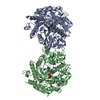
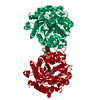
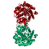
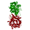
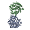
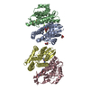
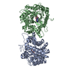
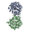
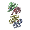
| Title | Crystal structure of the inactive mutant Monocot (Maize ZMGlu1) beta-glucosidase ZMGluE191D in complex with the competitive inhibitor dhurrin | ||||||
 Components Components | BETA-GLUCOSIDASE | ||||||
 Keywords Keywords | HYDROLASE / GLYCOSIDE HYDROLASE / BETA-GLUCOSIDASE / FAMILY 1 / RETENTION OF THE ANOMERIC CONFIGURATION / INACTIVE MUTANT E191D / COMPLEX WITH DHURRIN | ||||||
| Function / homology |  Function and homology informationfucosidase activity / xylanase activity / galactosidase activity / 4-hydroxy-7-methoxy-3-oxo-3,4-dihydro-2H-1,4-benzoxazin-2-yl glucoside beta-D-glucosidase / DIMBOA glucoside beta-D-glucosidase activity / cytokinin-activated signaling pathway / mannosidase activity / cellulose 1,4-beta-cellobiosidase activity / scopolin beta-glucosidase activity / beta-glucosidase activity ...fucosidase activity / xylanase activity / galactosidase activity / 4-hydroxy-7-methoxy-3-oxo-3,4-dihydro-2H-1,4-benzoxazin-2-yl glucoside beta-D-glucosidase / DIMBOA glucoside beta-D-glucosidase activity / cytokinin-activated signaling pathway / mannosidase activity / cellulose 1,4-beta-cellobiosidase activity / scopolin beta-glucosidase activity / beta-glucosidase activity / beta-glucosidase / chloroplast / carbohydrate metabolic process Function and homology informationfucosidase activity / xylanase activity / galactosidase activity / 4-hydroxy-7-methoxy-3-oxo-3,4-dihydro-2H-1,4-benzoxazin-2-yl glucoside beta-D-glucosidase / DIMBOA glucoside beta-D-glucosidase activity / cytokinin-activated signaling pathway / mannosidase activity / cellulose 1,4-beta-cellobiosidase activity / scopolin beta-glucosidase activity / beta-glucosidase activity ...fucosidase activity / xylanase activity / galactosidase activity / 4-hydroxy-7-methoxy-3-oxo-3,4-dihydro-2H-1,4-benzoxazin-2-yl glucoside beta-D-glucosidase / DIMBOA glucoside beta-D-glucosidase activity / cytokinin-activated signaling pathway / mannosidase activity / cellulose 1,4-beta-cellobiosidase activity / scopolin beta-glucosidase activity / beta-glucosidase activity / beta-glucosidase / chloroplast / carbohydrate metabolic processSimilarity search - Function | ||||||
| Biological species |  ZEA MAYS (maize) ZEA MAYS (maize) | ||||||
| Method | X-RAY DIFFRACTION / SYNCHROTRON / MOLECULAR REPLACEMENT / Resolution: 2 Å | ||||||
 Authors Authors | Czjzek, M. / Cicek, M. / Bevan, D.R. / Zamboni, V. / Henrissat, B. / Esen, A. | ||||||
 Citation Citation | Journal: Proc. Natl. Acad. Sci. U.S.A. / Year: 2000 Title: The mechanism of substrate (aglycone) specificity in beta-glucosidases is revealed by crystal structures of mutant maize beta-glucosidase-DIMBOA, -DIMBOAGlc, and -dhurrin complexes. Authors: Czjzek, M. / Cicek, M. / Zamboni, V. / Bevan, D.R. / Henrissat, B. / Esen, A. | ||||||
| History |
|
- Structure visualization
| Structure viewer | Molecule: MolmilJmol/JSmol |
|---|
- Downloads & links
-Download
| PDBx/mmCIF format | 1e55.cif.gz | 216.9 KB | Display | PDBx/mmCIF format |
|---|---|---|---|---|
| PDB format | pdb1e55.ent.gz | 173.1 KB | Display | PDB format |
| PDBx/mmJSON format | 1e55.json.gz | Tree view | PDBx/mmJSON format | |
| Others |  Other downloads Other downloads |
-Validation report
| Arichive directory | https://data.pdbj.org/pub/pdb/validation_reports/e5/1e55ftp://data.pdbj.org/pub/pdb/validation_reports/e5/1e55 | HTTPS FTP |
|---|
-Related structure data
| Related structure data | 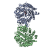 1e4lC  1e4nC  1e56C  1e1eS S: Starting model for refinement C: citing same article ( |
|---|---|
| Similar structure data |


-Links
 PDBj
PDBj


- Assembly
| Deposited unit | 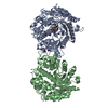
| ||||||||
|---|---|---|---|---|---|---|---|---|---|
| 1 |
| ||||||||
| Unit cell |
| ||||||||
| Noncrystallographic symmetry (NCS) | NCS oper: (Code: given Matrix: (-0.8236, 0.36629, -0.43302), Vector : |
-Components
| #1: Protein | Mass: 58458.422 Da / Num. of mol.: 2 / Mutation: YES Source method: isolated from a genetically manipulated source Source: (gene. exp.) ZEA MAYS (maize) / Strain: CV. MUTIN / Tissue: COLEOPTILE / Organelle: CHLOROPLAST / Plasmid: PET-21A / Gene (production host): GLU1 / Production host:  ESCHERICHIA COLI (E. coli) / Variant (production host): PLYSS / References: UniProt: P49235, beta-glucosidase ESCHERICHIA COLI (E. coli) / Variant (production host): PLYSS / References: UniProt: P49235, beta-glucosidase#2: Sugar | Glucose  Type: D-saccharide, beta linking / Mass: 180.156 Da / Num. of mol.: 2 Type: D-saccharide, beta linking / Mass: 180.156 Da / Num. of mol.: 2Source method: isolated from a genetically manipulated source Formula: C6H12O6 #3: Chemical |   Mass: 149.147 Da / Num. of mol.: 2 / Source method: obtained synthetically / Formula: C8H7NO2 Mass: 149.147 Da / Num. of mol.: 2 / Source method: obtained synthetically / Formula: C8H7NO2#4: Water | ChemComp-HOH / | Water Mass: 18.015 Da / Num. of mol.: 602 / Source method: isolated from a natural source / Formula: H2O Mass: 18.015 Da / Num. of mol.: 602 / Source method: isolated from a natural source / Formula: H2OCompound details | CHAIN A, B ENGINEERED | Sequence details | THE N- AND C-TERMINAL RESID. WERE NOT SEEN IN DENSITY MAPS | |
|---|
-Experimental details
-Experiment
| Experiment | Method: X-RAY DIFFRACTION / Number of used crystals: 1 |
|---|
- Sample preparation
| Crystal | Density Matthews: 2.21 Å3/Da / Density % sol: 44.43 % | ||||||||||||||||||||
|---|---|---|---|---|---|---|---|---|---|---|---|---|---|---|---|---|---|---|---|---|---|
| Crystal grow | pH: 7.5 / Details: 22 % PEG 4000, 5 % ISOPROPANOL, 0.1 M HEPES PH 7.5 | ||||||||||||||||||||
| Crystal grow | *PLUS Method: vapor diffusion, hanging drop | ||||||||||||||||||||
| Components of the solutions | *PLUS
|
-Data collection
| Diffraction | Mean temperature: 100 K |
|---|---|
| Diffraction source | Source: SYNCHROTRON / Site: ESRF  / Beamline: ID14-2 / Wavelength: 0.93 / Beamline: ID14-2 / Wavelength: 0.93 |
| Detector | Date: Mar 15, 2000 |
| Radiation | Protocol: SINGLE WAVELENGTH / Monochromatic (M) / Laue (L): M / Scattering type: x-ray |
| Radiation wavelength | Wavelength: 0.93 Å / Relative weight: 1 |
| Reflection | Resolution: 2→29.1 Å / Num. obs: 69098 / % possible obs: 98.8 % / Redundancy: 3 % / Rmerge(I) obs: 0.117 / Rsym value: 0.084 / Net I/σ(I): 6.3 |
| Reflection shell | Resolution: 2→2.11 Å / Redundancy: 2.8 % / Rmerge(I) obs: 0.397 / Mean I/σ(I) obs: 1.3 / Rsym value: 0.284 / % possible all: 97.1 |
| Reflection shell | *PLUS % possible obs: 97.1 % |
- Processing
| Software |
| ||||||||||||||||||||||||||||||||||||||||||||||||||||||||||||
|---|---|---|---|---|---|---|---|---|---|---|---|---|---|---|---|---|---|---|---|---|---|---|---|---|---|---|---|---|---|---|---|---|---|---|---|---|---|---|---|---|---|---|---|---|---|---|---|---|---|---|---|---|---|---|---|---|---|---|---|---|---|
| Refinement | Method to determine structure: MOLECULAR REPLACEMENT Starting model: 1E1E Resolution: 2→29 Å / Data cutoff high absF: 1000000 / Cross valid method: THROUGHOUT / σ(F): 0.05
| ||||||||||||||||||||||||||||||||||||||||||||||||||||||||||||
| Solvent computation | Solvent model: FLAT MODEL / Bsol: 41.17 Å2 / ksol: 0.3604 e/Å3 | ||||||||||||||||||||||||||||||||||||||||||||||||||||||||||||
| Displacement parameters | Biso mean: 26.1 Å2 | ||||||||||||||||||||||||||||||||||||||||||||||||||||||||||||
| Refine analyze |
| ||||||||||||||||||||||||||||||||||||||||||||||||||||||||||||
| Refinement step | Cycle: LAST / Resolution: 2→29 Å
| ||||||||||||||||||||||||||||||||||||||||||||||||||||||||||||
| Refine LS restraints |
| ||||||||||||||||||||||||||||||||||||||||||||||||||||||||||||
| Refine LS restraints NCS | Rms dev position: 0.274 Å / Weight position: 5 | ||||||||||||||||||||||||||||||||||||||||||||||||||||||||||||
| LS refinement shell | Resolution: 2→2.07 Å / Total num. of bins used: 10
| ||||||||||||||||||||||||||||||||||||||||||||||||||||||||||||
| Xplor file |
| ||||||||||||||||||||||||||||||||||||||||||||||||||||||||||||
| Software | *PLUS Name: CNS / Version: 1 / Classification: refinement | ||||||||||||||||||||||||||||||||||||||||||||||||||||||||||||
| Refine LS restraints | *PLUS
|
