 Movie
Movie Controller

[English] 日本語
 Yorodumi
- PDB-1e1h: Crystal Structure of recombinant Botulinum Neurotoxin Type A Ligh... -
+ Open data

- Basic information
| Entry | Database: PDB / ID: 1e1h | ||||||
|---|---|---|---|---|---|---|---|
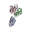
| Title | Crystal Structure of recombinant Botulinum Neurotoxin Type A Light Chain, self-inhibiting Zn endopeptidase. | ||||||
 Components Components | (BOTULINUM NEUROTOXIN TYPE A LIGHT CHAIN) x 2 | ||||||
 Keywords Keywords |  HYDROLASE / NEUROTOXIN / ZN-ENDOPEPTIDASE / COMPLEX / SUBSTRATE BOUND / BOTULINUM / INHIBITOR BOUND HYDROLASE / NEUROTOXIN / ZN-ENDOPEPTIDASE / COMPLEX / SUBSTRATE BOUND / BOTULINUM / INHIBITOR BOUND | ||||||
| Function / homology |  Function and homology informationbontoxilysin / host cell presynaptic membrane / host cell cytoplasmic vesicle / host cell cytosol / protein transmembrane transporter activity / metalloendopeptidase activity / toxin activity / host cell plasma membrane / proteolysis / zinc ion binding ...bontoxilysin / host cell presynaptic membrane / host cell cytoplasmic vesicle / host cell cytosol / protein transmembrane transporter activity / metalloendopeptidase activity / toxin activity / host cell plasma membrane / proteolysis / zinc ion binding / extracellular region / membrane Function and homology informationbontoxilysin / host cell presynaptic membrane / host cell cytoplasmic vesicle / host cell cytosol / protein transmembrane transporter activity / metalloendopeptidase activity / toxin activity / host cell plasma membrane / proteolysis / zinc ion binding ...bontoxilysin / host cell presynaptic membrane / host cell cytoplasmic vesicle / host cell cytosol / protein transmembrane transporter activity / metalloendopeptidase activity / toxin activity / host cell plasma membrane / proteolysis / zinc ion binding / extracellular region / membraneSimilarity search - Function | ||||||
| Biological species |   CLOSTRIDIUM BOTULINUM (bacteria) CLOSTRIDIUM BOTULINUM (bacteria) | ||||||
| Method | X-RAY DIFFRACTION / SYNCHROTRON / MOLECULAR REPLACEMENT / Resolution: 1.8 Å | ||||||
 Authors Authors | Knapp, M. / Rupp, B. | ||||||
 Citation Citation | Journal: Proc.Natl.Acad.Sci.USA / Year: 2004 Title: Crystal Structure of Clostridium Botulinum Neurotoxin Protease in a Product-Bound State: Evidence for Noncanonical Zinc Protease Activity Authors: Segelke, B.W. / Knapp, M. / Kadhkodayan, S. / Balhorn, R. / Rupp, B. | ||||||
| History |
|
- Structure visualization
| Structure viewer | Molecule: MolmilJmol/JSmol |
|---|
- Downloads & links
-Download
| PDBx/mmCIF format | 1e1h.cif.gz | 190.7 KB | Display | PDBx/mmCIF format |
|---|---|---|---|---|
| PDB format | pdb1e1h.ent.gz | 148.8 KB | Display | PDB format |
| PDBx/mmJSON format | 1e1h.json.gz | Tree view | PDBx/mmJSON format | |
| Others |  Other downloads Other downloads |
-Validation report
| Arichive directory | https://data.pdbj.org/pub/pdb/validation_reports/e1/1e1hftp://data.pdbj.org/pub/pdb/validation_reports/e1/1e1h | HTTPS FTP |
|---|
-Related structure data
| Related structure data |  3btaS S: Starting model for refinement |
|---|---|
| Similar structure data |


-Links
 PDBj
PDBj- Assembly
| Deposited unit | 
| ||||||||||||
|---|---|---|---|---|---|---|---|---|---|---|---|---|---|
| 1 |
| ||||||||||||
| Unit cell |
| ||||||||||||
| Noncrystallographic symmetry (NCS) | NCS oper:
|
-Components
| #1: Protein | Mass: 32153.004 Da / Num. of mol.: 2 / Fragment: RESIDUES 10-250 Source method: isolated from a genetically manipulated source Source: (gene. exp.) CLOSTRIDIUM BOTULINUM (bacteria) / Strain: KYOTO-F / Production host: ESCHERICHIA COLI (E. coli) / Variant (production host): BLR DE-3 / References: UniProt: Q45894, bontoxilysin#2: Protein | Mass: 20560.143 Da / Num. of mol.: 2 / Fragment: RESIDUES 252-416 / Mutation: YES Source method: isolated from a genetically manipulated source Details: HOMODIMER, CONTAINING CLEAVED SUBSTRATE ANALOG (LOOP 245-255) IN ACTIVE SITE Source: (gene. exp.) CLOSTRIDIUM BOTULINUM (bacteria) / Strain: KYOTO-F / Production host: ESCHERICHIA COLI (E. coli) / Variant (production host): BLR DE-3 / References: UniProt: Q45894, bontoxilysin#3: Chemical |   Mass: 65.409 Da / Num. of mol.: 2 / Source method: obtained synthetically / Formula: Zn Mass: 65.409 Da / Num. of mol.: 2 / Source method: obtained synthetically / Formula: Zn#4: Water | ChemComp-HOH / | Water Mass: 18.015 Da / Num. of mol.: 768 / Source method: isolated from a natural source / Formula: H2O Mass: 18.015 Da / Num. of mol.: 768 / Source method: isolated from a natural source / Formula: H2OCompound details | TER TYR: PEPTIDE CHAIN CLEAVED AT AA 249 AND 250. HIS: PEPTIDE CHAIN CLEAVED AT AA 249 AND 250. | Sequence details | EXPERIMENTAL PROTEIN HAS 6XHIS-TAG AND S-TAG AT N-TERMINUS FOLLOWED BY RESIDUES 9-415 OF NCBI: ...EXPERIMENT | |
|---|
-Experimental details
-Experiment
| Experiment | Method: X-RAY DIFFRACTION / Number of used crystals: 1 |
|---|
- Sample preparation
| Crystal | Density Matthews: 2.91 Å3/Da / Density % sol: 53 % | ||||||||||||||||||||||||||||||||||||||||||||||||||||||||||||
|---|---|---|---|---|---|---|---|---|---|---|---|---|---|---|---|---|---|---|---|---|---|---|---|---|---|---|---|---|---|---|---|---|---|---|---|---|---|---|---|---|---|---|---|---|---|---|---|---|---|---|---|---|---|---|---|---|---|---|---|---|---|
| Crystal grow | Method: vapor diffusion, hanging drop / pH: 4.6 Details: HANGING DROP VAPOUR DIFFUSION, DROP: 4UL 5MG/ML PROTEIN & 2UL WELL. PROTEIN: 0.05M TRIS PH 8.0,10% GLYCEROL, 0.1% TRITON X-100,1.0MM 2-ME,4% XYLITOL. WELL: 0.2M (NH4)2SO4,0.1M NAOAC PH 4.6, 25% PEG4000. | ||||||||||||||||||||||||||||||||||||||||||||||||||||||||||||
| Crystal grow | *PLUS Temperature: 22 ℃ / pH: 8 / Method: vapor diffusion, hanging drop | ||||||||||||||||||||||||||||||||||||||||||||||||||||||||||||
| Components of the solutions | *PLUS
|
-Data collection
| Diffraction | Mean temperature: 125 K |
|---|---|
| Diffraction source | Source: SYNCHROTRON / Site: ALS  / Beamline: 5.0.2 / Wavelength: 1.1 / Beamline: 5.0.2 / Wavelength: 1.1 |
| Detector | Type: ADSC CCD / Detector: CCD / Date: Jan 15, 2000 / Details: DOUBLE FOCUSSING |
| Radiation | Monochromator: SI / Protocol: SINGLE WAVELENGTH / Monochromatic (M) / Laue (L): M / Scattering type: x-ray |
| Radiation wavelength | Wavelength: 1.1 Å / Relative weight: 1 |
| Reflection | Resolution: 1.8→19.34 Å / Num. obs: 93168 / % possible obs: 95.2 % / Observed criterion σ(I): 0 / Redundancy: 1.9 % / Biso Wilson estimate: 24.47 Å2 / Rsym value: 0.042 / Net I/σ(I): 7.9 |
| Reflection shell | Resolution: 1.8→1.9 Å / Redundancy: 1.9 % / Mean I/σ(I) obs: 1.7 / Rsym value: 0.383 / % possible all: 95.2 |
| Reflection | *PLUS Highest resolution: 1.8 Å / Redundancy: 3.3 % / Rmerge(I) obs: 0.042 |
| Reflection shell | *PLUS Highest resolution: 1.8 Å / Lowest resolution: 1.9 Å / Redundancy: 1.9 % / Num. unique obs: 6918 / Rmerge(I) obs: 0.383 / Mean I/σ(I) obs: 1.7 |
- Processing
| Software |
| ||||||||||||||||||||||||||||||||||||||||||||||||||||||||||||||||||||||||||||||||||||
|---|---|---|---|---|---|---|---|---|---|---|---|---|---|---|---|---|---|---|---|---|---|---|---|---|---|---|---|---|---|---|---|---|---|---|---|---|---|---|---|---|---|---|---|---|---|---|---|---|---|---|---|---|---|---|---|---|---|---|---|---|---|---|---|---|---|---|---|---|---|---|---|---|---|---|---|---|---|---|---|---|---|---|---|---|---|
| Refinement | Method to determine structure: MOLECULAR REPLACEMENT Starting model: LC COORDINATES OF PDB ENTRY 3BTA Resolution: 1.8→19.34 Å / SU B: 2.746 / SU ML: 0.085 / Cross valid method: THROUGHOUT / σ(F): 0 / ESU R: 0.129 / ESU R Free: 0.127
| ||||||||||||||||||||||||||||||||||||||||||||||||||||||||||||||||||||||||||||||||||||
| Displacement parameters | Biso mean: 31.5 Å2 | ||||||||||||||||||||||||||||||||||||||||||||||||||||||||||||||||||||||||||||||||||||
| Refinement step | Cycle: LAST / Resolution: 1.8→19.34 Å
| ||||||||||||||||||||||||||||||||||||||||||||||||||||||||||||||||||||||||||||||||||||
| Refine LS restraints |
| ||||||||||||||||||||||||||||||||||||||||||||||||||||||||||||||||||||||||||||||||||||
| Refinement | *PLUS Highest resolution: 1.8 Å | ||||||||||||||||||||||||||||||||||||||||||||||||||||||||||||||||||||||||||||||||||||
| Solvent computation | *PLUS | ||||||||||||||||||||||||||||||||||||||||||||||||||||||||||||||||||||||||||||||||||||
| Displacement parameters | *PLUS | ||||||||||||||||||||||||||||||||||||||||||||||||||||||||||||||||||||||||||||||||||||
| Refine LS restraints | *PLUS
|
