 Movie
Movie Controller


+ Open data

- Basic information
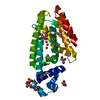
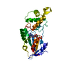
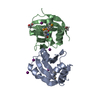
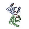
| Entry | Database: PDB / ID: 1dz1 | ||||||
|---|---|---|---|---|---|---|---|
| Title | Mouse HP1 (M31) C terminal (shadow chromo) domain | ||||||
 Components Components | MODIFIER 1 PROTEIN | ||||||
 Keywords Keywords |  CHROMATIN STRUCTURE / CHROMO DOMAIN / HETEROCHROMATIN PROTEIN PROTEIN INTERACTION / DIMERIC CHROMATIN STRUCTURE / CHROMO DOMAIN / HETEROCHROMATIN PROTEIN PROTEIN INTERACTION / DIMERIC | ||||||
| Function / homology |  Function and homology information Function and homology informationchromocenter / histone methyltransferase binding / male pronucleus / female pronucleus / site of DNA damage / chromosome, centromeric region / pericentric heterochromatin / methylated histone binding / spindle / chromatin organization ...chromocenter / histone methyltransferase binding / male pronucleus / female pronucleus / site of DNA damage / chromosome, centromeric region / pericentric heterochromatin / methylated histone binding / spindle / chromatin organization / chromosome, telomeric region / molecular adaptor activity / nuclear body / intracellular membrane-bounded organelle / negative regulation of DNA-templated transcription / DNA damage response / chromatin binding / chromatin / negative regulation of transcription by RNA polymerase II / enzyme binding / DNA binding / nucleoplasm / identical protein binding / nucleusSimilarity search - Function | ||||||
| Biological species |  MUS MUSCULUS (house mouse) MUS MUSCULUS (house mouse) | ||||||
| Method | SOLUTION NMR | ||||||
 Authors Authors | Brasher, S.V. / Smith, B.O. / Fogh, R.H. / Nietlispach, D. / Thiru, A. / Nielsen, P.R. / Broadhurst, R.W. / Ball, L.J. / Murzina, N. / Laue, E.D. | ||||||
 Citation Citation | Journal: Embo J. / Year: 2000 Title: The Structure of Mouse Hp1 Suggests a Unique Mode of Single Peptide Recognition by the Shadow Chromo Domain Dimer Authors: Brasher, S.V. / Smith, B.O. / Fogh, R.H. / Nietlispach, D. / Thiru, A. / Nielsen, P.R. / Broadhurst, R.W. / Ball, L.J. / Murzina, N. / Laue, E.D. | ||||||
| History |
|
- Structure visualization
| Structure viewer | Molecule: MolmilJmol/JSmol |
|---|
- Downloads & links
-Download
| PDBx/mmCIF format | 1dz1.cif.gz | 695.2 KB | Display | PDBx/mmCIF format |
|---|---|---|---|---|
| PDB format | pdb1dz1.ent.gz | 602.5 KB | Display | PDB format |
| PDBx/mmJSON format | 1dz1.json.gz | Tree view | PDBx/mmJSON format | |
| Others |  Other downloads Other downloads |
-Validation report
| Arichive directory | https://data.pdbj.org/pub/pdb/validation_reports/dz/1dz1ftp://data.pdbj.org/pub/pdb/validation_reports/dz/1dz1 | HTTPS FTP |
|---|
-Related structure data
| Related structure data | |
|---|---|
| Similar structure data |


-Links
 PDBj
PDBj- Assembly
| Deposited unit | 
| |||||||||
|---|---|---|---|---|---|---|---|---|---|---|
| 1 |
| |||||||||
| NMR ensembles |
|
-Components
| #1: Protein | Mass: 8076.179 Da / Num. of mol.: 2 / Fragment: SHADOW CHROMO DOMAIN, RESIDUES 104-171 Source method: isolated from a genetically manipulated source Source: (gene. exp.) MUS MUSCULUS (house mouse) / Plasmid: PET16B / Production host: Escherichia coli BL21(DE3) / References: UniProt: P83917Sequence details | HIS A 102 CLONING ARTIFACT MUTATION ARISING FROM VECTOR MET A 103 CLONING ARTIFACT MUTATION ARISING ...HIS A 102 CLONING ARTIFACT MUTATION ARISING FROM VECTOR MET A 103 CLONING ARTIFACT MUTATION ARISING FROM VECTOR HIS B 102 CLONING ARTIFACT MUTATION ARISING FROM VECTOR MET B 103 CLONING ARTIFACT MUTATION ARISING FROM VECTOR | |
|---|
-Experimental details
-Experiment
| Experiment | Method: SOLUTION NMR |
|---|---|
| NMR details | Text: THE STRUCTURE WAS DETERMINED USING TRIPLE-RESONANCE NMR SPECTROSCOPY ON 13C, 15N-LABELED PROTEIN. DOUBLE-HALF FILTERED 2D NOESY AND HALF-FILTERED HSQC-NOESY ON A MIXED LABELED/UNLABELED SAMPLE ...Text: THE STRUCTURE WAS DETERMINED USING TRIPLE-RESONANCE NMR SPECTROSCOPY ON 13C, 15N-LABELED PROTEIN. DOUBLE-HALF FILTERED 2D NOESY AND HALF-FILTERED HSQC-NOESY ON A MIXED LABELED/UNLABELED SAMPLE WERE USED TO DISTINGUISH INTRA- AND INTER-MONOMER NOES. RESIDUES 102-109 AND 171 ARE INSUFFICIENTLY CONSTRAINED BY THE INPUT DATA AND SHOULD BE CONSIDERED OF UNKNOWN STRUCTURE. THUS THE STRUCTURE IS ONLY DEFINED FOR RESIDUES 110 - 170. |
- Sample preparation
| Details | Contents: 0.6-1.4 MM MOUSE HP1 C-TERMINAL DOMAIN (AS MONOMER), 10 MM SODIUM PHOSPHATE BUFFER, 10 MM PERDEUTERATED DTT, 0.05% SODIUM AZIDE |
|---|---|
| Sample conditions | pH: 8.0 / Pressure: 1 atm / Temperature: 303 K |
| Crystal grow | *PLUS Method: other / Details: NMR |
-NMR measurement
| NMR spectrometer |
|
|---|
- Processing
| NMR software |
| ||||||||||||||||||||||||
|---|---|---|---|---|---|---|---|---|---|---|---|---|---|---|---|---|---|---|---|---|---|---|---|---|---|
| Refinement | Software ordinal: 1 Details: REFINEMENT DETAILS CAN BE FOUND IN THE JRNL CITATION ABOVE. STRUCTURE WAS GENERATED FROM EXPERIMENTS CARRIED OUT AT 303-308K WITH PH 7.5-8.0 | ||||||||||||||||||||||||
| NMR ensemble | Conformer selection criteria: LEAST NOE VIOLATION ENERGY / Conformers calculated total number: 35 / Conformers submitted total number: 16 |
