 Movie
Movie Controller

[English] 日本語
 Yorodumi
- PDB-1dwo: Crystal Structure of Hydroxynitrile Lyase from Manihot esculenta ... -
+ Open data

- Basic information
| Entry | Database: PDB / ID: 1dwo | ||||||
|---|---|---|---|---|---|---|---|
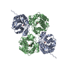
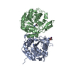
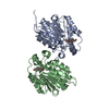
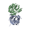
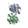
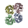
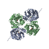
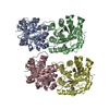
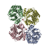
| Title | Crystal Structure of Hydroxynitrile Lyase from Manihot esculenta in Complex with Substrates Acetone and Chloroacetone:Implications for the Mechanism of Cyanogenesis | ||||||
 Components Components | HYDROXYNITRILE LYASE | ||||||
 Keywords Keywords | HYDROXYNITRILE LYASE /  ACETONE COMPLEX ACETONE COMPLEX | ||||||
| Function / homology |  Function and homology information(S)-hydroxynitrile lyase / aliphatic (S)-hydroxynitrile lyase activity / aromatic (S)-hydroxynitrile lyase activity Function and homology information(S)-hydroxynitrile lyase / aliphatic (S)-hydroxynitrile lyase activity / aromatic (S)-hydroxynitrile lyase activitySimilarity search - Function | ||||||
| Biological species |  MANIHOT ESCULENTA (cassava) MANIHOT ESCULENTA (cassava) | ||||||
| Method | X-RAY DIFFRACTION / SYNCHROTRON / MOLECULAR REPLACEMENT / Resolution: 2.2 Å | ||||||
 Authors Authors | Lauble, H. / Forster, S. / Miehlich, B. / Wajant, H. / Effenberger, F. | ||||||
 Citation Citation | Journal: Acta Crystallogr.,Sect.D / Year: 2001 Title: Structure of Hydroxynitrile Lyase from Manihot Esculenta in Complex with Substrates Acetone and Chloroacetone: Implications for the Mechanism of Cyanogenesis Authors: Lauble, H. / Forster, S. / Miehlich, B. / Wajant, H. / Effenberger, F. | ||||||
| History |
|
- Structure visualization
| Structure viewer | Molecule: MolmilJmol/JSmol |
|---|
- Downloads & links
-Download
| PDBx/mmCIF format | 1dwo.cif.gz | 115.8 KB | Display | PDBx/mmCIF format |
|---|---|---|---|---|
| PDB format | pdb1dwo.ent.gz | 95 KB | Display | PDB format |
| PDBx/mmJSON format | 1dwo.json.gz | Tree view | PDBx/mmJSON format | |
| Others |  Other downloads Other downloads |
-Validation report
| Arichive directory | https://data.pdbj.org/pub/pdb/validation_reports/dw/1dwoftp://data.pdbj.org/pub/pdb/validation_reports/dw/1dwo | HTTPS FTP |
|---|
-Related structure data

-Links
 PDBj
PDBj


- Assembly
| Deposited unit | 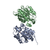
| ||||||||
|---|---|---|---|---|---|---|---|---|---|
| 1 | 
| ||||||||
| Unit cell |
| ||||||||
| Details | BIOLOGICAL_UNIT: TETRAMER |
-Components
| #1: Protein | Mass: 29835.209 Da / Num. of mol.: 2 Source method: isolated from a genetically manipulated source Details: ACETONE COMPLEX / Source: (gene. exp.) MANIHOT ESCULENTA (cassava) / Description: RECOMBINANT PROTEIN / Production host:  ESCHERICHIA COLI (E. coli) / References: UniProt: P52705, EC: 4.2.1.37 ESCHERICHIA COLI (E. coli) / References: UniProt: P52705, EC: 4.2.1.37#2: Chemical | ChemComp-ACN / | Acetone  Mass: 58.079 Da / Num. of mol.: 1 / Source method: obtained synthetically / Formula: C3H6O Mass: 58.079 Da / Num. of mol.: 1 / Source method: obtained synthetically / Formula: C3H6O#3: Water | ChemComp-HOH / | Water Mass: 18.015 Da / Num. of mol.: 271 / Source method: isolated from a natural source / Formula: H2O Mass: 18.015 Da / Num. of mol.: 271 / Source method: isolated from a natural source / Formula: H2OSequence details | THE SWISSPROT ENTRY P52705 REPORTS THIS ENZYME TO BE EC 4.2.1.39, AND TO HAVE A HOMOTRIMERIC ...THE SWISSPROT ENTRY P52705 REPORTS THIS ENZYME TO BE EC 4.2.1.39, AND TO HAVE A HOMOTRIMER | |
|---|
-Experimental details
-Experiment
| Experiment | Method: X-RAY DIFFRACTION / Number of used crystals: 1 |
|---|
- Sample preparation
| Crystal | Density Matthews: 4.3 Å3/Da / Density % sol: 71 % | |||||||||||||||||||||||||
|---|---|---|---|---|---|---|---|---|---|---|---|---|---|---|---|---|---|---|---|---|---|---|---|---|---|---|
| Crystal grow | pH: 5.4 / Details: 0.1 M NACITRAT, PH 5.4, 6% PEG8000, 28% MPD | |||||||||||||||||||||||||
| Crystal grow | *PLUS Method: vapor diffusion, hanging drop / Details: Lauble, H., (1999) Acta Crystallogr., D55, 904. | |||||||||||||||||||||||||
| Components of the solutions | *PLUS
|
-Data collection
| Diffraction | Mean temperature: 100 K |
|---|---|
| Diffraction source | Source: SYNCHROTRON / Site: EMBL/DESY, HAMBURG  / Beamline: X11 / Wavelength: 0.905 / Beamline: X11 / Wavelength: 0.905 |
| Detector | Type: MARRESEARCH / Detector: IMAGE PLATE |
| Radiation | Protocol: SINGLE WAVELENGTH / Monochromatic (M) / Laue (L): M / Scattering type: x-ray |
| Radiation wavelength | Wavelength: 0.905 Å / Relative weight: 1 |
| Reflection | Resolution: 2.2→20 Å / Num. obs: 52853 / % possible obs: 97.1 % / Redundancy: 4.2 % / Rmerge(I) obs: 0.054 / Rsym value: 0.068 / Net I/σ(I): 8.6 |
| Reflection shell | Resolution: 2.2→2.3 Å / Redundancy: 4.1 % / Mean I/σ(I) obs: 3.2 / % possible all: 99.4 |
| Reflection | *PLUS Lowest resolution: 15 Å / Num. measured all: 215742 / Rmerge(I) obs: 0.068 |
| Reflection shell | *PLUS % possible obs: 99.4 % / Rmerge(I) obs: 0.141 |
- Processing
| Software |
| ||||||||||||||||||||||||||||||||||||||||||||||||||||||||||||
|---|---|---|---|---|---|---|---|---|---|---|---|---|---|---|---|---|---|---|---|---|---|---|---|---|---|---|---|---|---|---|---|---|---|---|---|---|---|---|---|---|---|---|---|---|---|---|---|---|---|---|---|---|---|---|---|---|---|---|---|---|---|
| Refinement | Method to determine structure: MOLECULAR REPLACEMENT / Resolution: 2.2→8 Å / Data cutoff high absF: 10000000 / Data cutoff low absF: 0.001 / σ(F): 2
| ||||||||||||||||||||||||||||||||||||||||||||||||||||||||||||
| Displacement parameters | Biso mean: 26 Å2 | ||||||||||||||||||||||||||||||||||||||||||||||||||||||||||||
| Refine analyze |
| ||||||||||||||||||||||||||||||||||||||||||||||||||||||||||||
| Refinement step | Cycle: LAST / Resolution: 2.2→8 Å
| ||||||||||||||||||||||||||||||||||||||||||||||||||||||||||||
| Refine LS restraints |
| ||||||||||||||||||||||||||||||||||||||||||||||||||||||||||||
| Xplor file | Serial no: 1 / Param file: PARAM19X.PRO / Topol file: TOPH19X.PRO |
