 Movie
Movie Controller

[English] 日本語
 Yorodumi
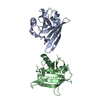
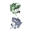
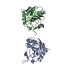
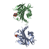
- PDB-1dvk: CRYSTAL STRUCTURE OF THE FUNCTIONAL DOMAIN OF THE SPLICING FACTOR... -
+ Open data

- Basic information
| Entry | Database: PDB / ID: 1dvk | ||||||
|---|---|---|---|---|---|---|---|
| Title | CRYSTAL STRUCTURE OF THE FUNCTIONAL DOMAIN OF THE SPLICING FACTOR PRP18 | ||||||
 Components Components | PRP18 | ||||||
 Keywords Keywords |  RNA BINDING PROTEIN / pre-mRNA Splicing factor / Prp18 RNA BINDING PROTEIN / pre-mRNA Splicing factor / Prp18 | ||||||
| Function / homology |  Function and homology information Function and homology informationU2-type post-spliceosomal complex / nuclear mRNA surveillance / generation of catalytic spliceosome for second transesterification step / U5 snRNP / U4/U6 x U5 tri-snRNP complex / mRNA splicing, via spliceosomeSimilarity search - Function | ||||||
| Biological species |  Saccharomyces cerevisiae (brewer's yeast) Saccharomyces cerevisiae (brewer's yeast) | ||||||
| Method | X-RAY DIFFRACTION / SYNCHROTRON / Resolution: 2.15 Å | ||||||
 Authors Authors | Jiang, J. / Horowitz, D.S. / Xu, R.M. | ||||||
 Citation Citation | Journal: Proc.Natl.Acad.Sci.USA / Year: 2000 Title: Crystal structure of the functional domain of the splicing factor Prp18. Authors: Jiang, J. / Horowitz, D.S. / Xu, R.M. | ||||||
| History |
|
- Structure visualization
| Structure viewer | Molecule: MolmilJmol/JSmol |
|---|
- Downloads & links
-Download
| PDBx/mmCIF format | 1dvk.cif.gz | 75.1 KB | Display | PDBx/mmCIF format |
|---|---|---|---|---|
| PDB format | pdb1dvk.ent.gz | 57.1 KB | Display | PDB format |
| PDBx/mmJSON format | 1dvk.json.gz | Tree view | PDBx/mmJSON format | |
| Others |  Other downloads Other downloads |
-Validation report
| Arichive directory | https://data.pdbj.org/pub/pdb/validation_reports/dv/1dvkftp://data.pdbj.org/pub/pdb/validation_reports/dv/1dvk | HTTPS FTP |
|---|
-Related structure data
| Similar structure data |
|---|


-Links
 PDBj
PDBj- Assembly
| Deposited unit | 
| ||||||||
|---|---|---|---|---|---|---|---|---|---|
| 1 |
| ||||||||
| Unit cell |
|
-Components
| #1: Protein | Mass: 19625.801 Da / Num. of mol.: 2 Source method: isolated from a genetically manipulated source Source: (gene. exp.) Saccharomyces cerevisiae (brewer's yeast)Species (production host): Escherichia coli / Production host:  Escherichia coli BL21(DE3) (bacteria) / Strain (production host): BL21(DE3) / References: UniProt: P33411 Escherichia coli BL21(DE3) (bacteria) / Strain (production host): BL21(DE3) / References: UniProt: P33411#2: Water | ChemComp-HOH / | Water Mass: 18.015 Da / Num. of mol.: 219 / Source method: isolated from a natural source / Formula: H2O Mass: 18.015 Da / Num. of mol.: 219 / Source method: isolated from a natural source / Formula: H2O |
|---|
-Experimental details
-Experiment
| Experiment | Method: X-RAY DIFFRACTION / Number of used crystals: 1 |
|---|
- Sample preparation
| Crystal | Density Matthews: 2.15 Å3/Da / Density % sol: 42.91 % | ||||||||||||||||||||||||||||||
|---|---|---|---|---|---|---|---|---|---|---|---|---|---|---|---|---|---|---|---|---|---|---|---|---|---|---|---|---|---|---|---|
| Crystal grow | Temperature: 290 K / Method: vapor diffusion, hanging drop / pH: 6 Details: 100mM MES 6.0, 25% PEG8000, 10% Glycerol, VAPOR DIFFUSION, HANGING DROP, temperature 290K | ||||||||||||||||||||||||||||||
| Crystal grow | *PLUS Temperature: 17 ℃ | ||||||||||||||||||||||||||||||
| Components of the solutions | *PLUS
|
-Data collection
| Diffraction | Mean temperature: 100 K |
|---|---|
| Diffraction source | Source: SYNCHROTRON / Site: NSLS  / Beamline: X26C / Wavelength: 1.09 / Beamline: X26C / Wavelength: 1.09 |
| Detector | Type: FUJI / Detector: IMAGE PLATE / Date: Oct 10, 1998 |
| Radiation | Protocol: SINGLE WAVELENGTH / Monochromatic (M) / Laue (L): M / Scattering type: x-ray |
| Radiation wavelength | Wavelength: 1.09 Å / Relative weight: 1 |
| Reflection | Resolution: 2.15→40 Å / Num. all: 18919 / Num. obs: 17664 / % possible obs: 93.2 % / Observed criterion σ(F): 0 / Observed criterion σ(I): 0 / Redundancy: 10.7 % / Biso Wilson estimate: 19.59 Å2 / Rmerge(I) obs: 0.041 / Net I/σ(I): 16.9 |
| Reflection shell | Resolution: 2.15→2.23 Å / Redundancy: 4.5 % / Rmerge(I) obs: 0.097 / % possible all: 55.7 |
| Reflection | *PLUS Num. measured all: 188477 |
- Processing
| Software |
| |||||||||||||||||||||||||
|---|---|---|---|---|---|---|---|---|---|---|---|---|---|---|---|---|---|---|---|---|---|---|---|---|---|---|
| Refinement | Resolution: 2.15→40 Å / σ(F): 0 / σ(I): 0 / Stereochemistry target values: Engh & Huber
| |||||||||||||||||||||||||
| Refinement step | Cycle: LAST / Resolution: 2.15→40 Å
| |||||||||||||||||||||||||
| Refine LS restraints |
| |||||||||||||||||||||||||
| Software | *PLUS Name: CNS / Classification: refinement | |||||||||||||||||||||||||
| Refinement | *PLUS Lowest resolution: 40 Å / σ(F): 0 | |||||||||||||||||||||||||
| Solvent computation | *PLUS | |||||||||||||||||||||||||
| Displacement parameters | *PLUS | |||||||||||||||||||||||||
| Refine LS restraints | *PLUS
|
