 Movie
Movie Controller

[English] 日本語
 Yorodumi
- PDB-1dvf: IDIOTOPIC ANTIBODY D1.3 FV FRAGMENT-ANTIIDIOTOPIC ANTIBODY E5.2 F... -
+ Open data

- Basic information
| Entry | Database: PDB / ID: 1dvf | ||||||
|---|---|---|---|---|---|---|---|
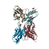
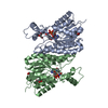
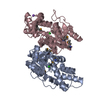
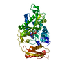
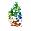
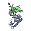
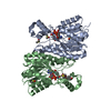
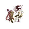
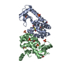
| Title | IDIOTOPIC ANTIBODY D1.3 FV FRAGMENT-ANTIIDIOTOPIC ANTIBODY E5.2 FV FRAGMENT COMPLEX | ||||||
 Components Components |
| ||||||
 Keywords Keywords | COMPLEX (IDIOTOPE-ANTIIDIOTOPE) /  IMMUNOGLOBULIN IMMUNOGLOBULIN | ||||||
| Function / homology |  Function and homology informationimmunoglobulin complex / immunoglobulin mediated immune response / antigen binding / adaptive immune response / immune response / extracellular space Function and homology informationimmunoglobulin complex / immunoglobulin mediated immune response / antigen binding / adaptive immune response / immune response / extracellular spaceSimilarity search - Function | ||||||
| Biological species |  Mus musculus (house mouse) Mus musculus (house mouse) | ||||||
| Method | X-RAY DIFFRACTION / Resolution: 1.9 Å | ||||||
 Authors Authors | Braden, B.C. / Fields, B.A. / Ysern, X. / Dall'Acqua, W. / Goldbaum, F.A. / Poljak, R.J. / Mariuzza, R.A. | ||||||
 Citation Citation | Journal: J.Mol.Biol. / Year: 1996 Title: Crystal structure of an Fv-Fv idiotope-anti-idiotope complex at 1.9 A resolution. Authors: Braden, B.C. / Fields, B.A. / Ysern, X. / Dall'Acqua, W. / Goldbaum, F.A. / Poljak, R.J. / Mariuzza, R.A. #1: Journal: Nature / Year: 1995Title: Molecular Basis of Antigen Mimicry by an Anti-Idiotope Authors: Fields, B.A. / Goldbaum, F.A. / Ysern, X. / Poljak, R.J. / Mariuzza, R.A. #2: Journal: J.Mol.Biol. / Year: 1994Title: Crystallization and Preliminary X-Ray Diffraction Study of an Idiotope-Anti-Idiotope Fv-Fv Complex Authors: Goldbaum, F.A. / Fields, B.A. / Cauerhff, A. / Ysern, X. / Houdusse, A. / Eisele, J.L. / Poljak, R.J. / Mariuzza, R.A. | ||||||
| History |
|
- Structure visualization
| Structure viewer | Molecule: MolmilJmol/JSmol |
|---|
- Downloads & links
-Download
| PDBx/mmCIF format | 1dvf.cif.gz | 99.9 KB | Display | PDBx/mmCIF format |
|---|---|---|---|---|
| PDB format | pdb1dvf.ent.gz | 80.5 KB | Display | PDB format |
| PDBx/mmJSON format | 1dvf.json.gz | Tree view | PDBx/mmJSON format | |
| Others |  Other downloads Other downloads |
-Validation report
| Arichive directory | https://data.pdbj.org/pub/pdb/validation_reports/dv/1dvfftp://data.pdbj.org/pub/pdb/validation_reports/dv/1dvf | HTTPS FTP |
|---|
-Related structure data
| Similar structure data |
|---|

-Links
 PDBj
PDBj

- Assembly
| Deposited unit | 
| ||||||||
|---|---|---|---|---|---|---|---|---|---|
| 1 |
| ||||||||
| 2 | 
| ||||||||
| Unit cell |
| ||||||||
| Components on special symmetry positions |
|
-Components
-Antibody , 4 types, 4 molecules ABCD
| #1: Antibody | Mass: 11858.171 Da / Num. of mol.: 1 Source method: isolated from a genetically manipulated source Details: IGG1, KAPPA / Source: (gene. exp.) Mus musculus (house mouse) / Strain: BALB-C / Production host:  Escherichia coli (E. coli) / References: UniProt: P01635 Escherichia coli (E. coli) / References: UniProt: P01635 |
|---|---|
| #2: Antibody | Mass: 12857.275 Da / Num. of mol.: 1 Source method: isolated from a genetically manipulated source Details: IGG1, KAPPA / Source: (gene. exp.) Mus musculus (house mouse) / Strain: BALB/C / Production host: Escherichia coli (E. coli) / References: UniProt: P01820 |
| #3: Antibody | Mass: 11770.961 Da / Num. of mol.: 1 Source method: isolated from a genetically manipulated source Details: IGG1, KAPPA / Source: (gene. exp.) Mus musculus (house mouse) / Strain: BALB-C / Production host: Escherichia coli (E. coli) / References: UniProt: P01646 |
| #4: Antibody | Mass: 13320.795 Da / Num. of mol.: 1 Source method: isolated from a genetically manipulated source Details: IGG1, KAPPA / Source: (gene. exp.) Mus musculus (house mouse) / Strain: BALB/C / Production host: Escherichia coli (E. coli) / References: UniProt: A0NA69*PLUS |
-Non-polymers , 2 types, 160 molecules 

| #5: Chemical |  Mass: 65.409 Da / Num. of mol.: 3 / Source method: obtained synthetically / Formula: Zn Mass: 65.409 Da / Num. of mol.: 3 / Source method: obtained synthetically / Formula: Zn#6: Water | ChemComp-HOH / | WaterMass: 18.015 Da / Num. of mol.: 157 / Source method: isolated from a natural source / Formula: H2O |
|---|
-Experimental details
-Experiment
| Experiment | Method: X-RAY DIFFRACTION |
|---|
- Sample preparation
| Crystal | Density Matthews: 3.09 Å3/Da / Density % sol: 60.21 % | |||||||||||||||||||||||||
|---|---|---|---|---|---|---|---|---|---|---|---|---|---|---|---|---|---|---|---|---|---|---|---|---|---|---|
| Crystal grow | *PLUS Method: vapor diffusion, hanging drop / Details: Goldbaum, F.A., (1994) J.Mol.Biol., 241, 739. | |||||||||||||||||||||||||
| Components of the solutions | *PLUS
|
-Data collection
| Diffraction source | Wavelength: 1.5418 |
|---|---|
| Detector | Type: RIGAKU / Detector: IMAGE PLATE |
| Radiation | Monochromatic (M) / Laue (L): M / Scattering type: x-ray |
| Radiation wavelength | Wavelength: 1.5418 Å / Relative weight: 1 |
| Reflection | Num. obs: 41404 / % possible obs: 82.5 % / Redundancy: 2.6 % / Rmerge(I) obs: 0.085 |
| Reflection | *PLUS Highest resolution: 1.9 Å / Num. measured all: 105969 |
- Processing
| Software |
| ||||||||||||||||||||||||||||||||||||||||||||||||||||||||||||
|---|---|---|---|---|---|---|---|---|---|---|---|---|---|---|---|---|---|---|---|---|---|---|---|---|---|---|---|---|---|---|---|---|---|---|---|---|---|---|---|---|---|---|---|---|---|---|---|---|---|---|---|---|---|---|---|---|---|---|---|---|---|
| Refinement | Resolution: 1.9→7 Å / σ(F): 2
| ||||||||||||||||||||||||||||||||||||||||||||||||||||||||||||
| Refinement step | Cycle: LAST / Resolution: 1.9→7 Å
| ||||||||||||||||||||||||||||||||||||||||||||||||||||||||||||
| Refine LS restraints |
| ||||||||||||||||||||||||||||||||||||||||||||||||||||||||||||
| Software | *PLUS Name: X-PLOR / Classification: refinement | ||||||||||||||||||||||||||||||||||||||||||||||||||||||||||||
| Refinement | *PLUS Rfactor Rfree: 0.235 | ||||||||||||||||||||||||||||||||||||||||||||||||||||||||||||
| Solvent computation | *PLUS | ||||||||||||||||||||||||||||||||||||||||||||||||||||||||||||
| Displacement parameters | *PLUS |
