 Movie
Movie Controller

[English] 日本語
 Yorodumi
- PDB-1dux: ELK-1/DNA STRUCTURE REVEALS HOW RESIDUES DISTAL FROM DNA-BINDING ... -
+ Open data

- Basic information
| Entry | Database: PDB / ID: 1dux | ||||||
|---|---|---|---|---|---|---|---|
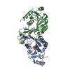
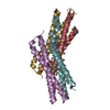
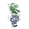
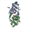
| Title | ELK-1/DNA STRUCTURE REVEALS HOW RESIDUES DISTAL FROM DNA-BINDING SURFACE AFFECT DNA-RECOGNITION | ||||||
 Components Components |
| ||||||
 Keywords Keywords | Transcription/DNA / ETS-DOMAIN /  DNA-BINDING DOMAIN / WINGED HELIX-TURN-HELIX / DNA-BINDING SPECIFICITY / Transcription-DNA COMPLEX DNA-BINDING DOMAIN / WINGED HELIX-TURN-HELIX / DNA-BINDING SPECIFICITY / Transcription-DNA COMPLEX | ||||||
| Function / homology |  Function and homology information Function and homology informationhippocampal neuron apoptotic process / response to fibroblast growth factor / cellular response to testosterone stimulus / NGF-stimulated transcription / ERK/MAPK targets / response to light stimulus / Estrogen-dependent nuclear events downstream of ESR-membrane signaling / axon terminus / nuclear receptor coactivator activity / liver development ...hippocampal neuron apoptotic process / response to fibroblast growth factor / cellular response to testosterone stimulus / NGF-stimulated transcription / ERK/MAPK targets / response to light stimulus / Estrogen-dependent nuclear events downstream of ESR-membrane signaling / axon terminus / nuclear receptor coactivator activity / liver development / mitochondrial membrane / lung development / cellular response to gamma radiation / HCMV Early Events / sequence-specific double-stranded DNA binding / gene expression / DNA-binding transcription activator activity, RNA polymerase II-specific / RNA polymerase II-specific DNA-binding transcription factor binding / transcription cis-regulatory region binding / DNA-binding transcription factor activity, RNA polymerase II-specific / RNA polymerase II cis-regulatory region sequence-specific DNA binding / DNA-binding transcription factor activity / dendrite / neuronal cell body / chromatin binding / chromatin / regulation of transcription by RNA polymerase II / positive regulation of DNA-templated transcription / positive regulation of transcription by RNA polymerase II / nucleoplasm / nucleusSimilarity search - Function | ||||||
| Biological species |  Homo sapiens (human) Homo sapiens (human) | ||||||
| Method | X-RAY DIFFRACTION / SYNCHROTRON / Resolution: 2.1 Å | ||||||
 Authors Authors | Mo, Y. / Vaessen, B. / Johnston, K. / Marmorstein, R. | ||||||
 Citation Citation | Journal: Nat.Struct.Biol. / Year: 2000 Title: Structure of the elk-1-DNA complex reveals how DNA-distal residues affect ETS domain recognition of DNA. Authors: Mo, Y. / Vaessen, B. / Johnston, K. / Marmorstein, R. | ||||||
| History |
|
- Structure visualization
| Structure viewer | Molecule: MolmilJmol/JSmol |
|---|
- Downloads & links
-Download
| PDBx/mmCIF format | 1dux.cif.gz | 82.4 KB | Display | PDBx/mmCIF format |
|---|---|---|---|---|
| PDB format | pdb1dux.ent.gz | 58.9 KB | Display | PDB format |
| PDBx/mmJSON format | 1dux.json.gz | Tree view | PDBx/mmJSON format | |
| Others |  Other downloads Other downloads |
-Validation report
| Arichive directory | https://data.pdbj.org/pub/pdb/validation_reports/du/1duxftp://data.pdbj.org/pub/pdb/validation_reports/du/1dux | HTTPS FTP |
|---|
-Related structure data
| Similar structure data |
|---|


-Links
 PDBj
PDBj


- Assembly
| Deposited unit | 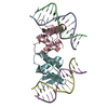
| ||||||||||
|---|---|---|---|---|---|---|---|---|---|---|---|
| 1 |
| ||||||||||
| Unit cell |
|
-Components
| #1: DNA chain | Mass: 4031.634 Da / Num. of mol.: 2 / Source method: obtained synthetically #2: DNA chain | Mass: 3911.562 Da / Num. of mol.: 2 / Source method: obtained synthetically #3: Protein | Mass: 11067.706 Da / Num. of mol.: 2 / Fragment: RESIDUES 1-94 Source method: isolated from a genetically manipulated source Source: (gene. exp.) Homo sapiens (human) / Plasmid: PRSETA / Production host:  Escherichia coli (E. coli) / References: UniProt: P19419 Escherichia coli (E. coli) / References: UniProt: P19419#4: Water | ChemComp-HOH / | Water Mass: 18.015 Da / Num. of mol.: 225 / Source method: isolated from a natural source / Formula: H2O Mass: 18.015 Da / Num. of mol.: 225 / Source method: isolated from a natural source / Formula: H2O |
|---|
-Experimental details
-Experiment
| Experiment | Method: X-RAY DIFFRACTION / Number of used crystals: 1 |
|---|
- Sample preparation
| Crystal | Density Matthews: 2.11 Å3/Da / Density % sol: 41.58 % | ||||||||||||||||||||||||||||||||||||||||||||||||||||||||||||||||||||||||||||||
|---|---|---|---|---|---|---|---|---|---|---|---|---|---|---|---|---|---|---|---|---|---|---|---|---|---|---|---|---|---|---|---|---|---|---|---|---|---|---|---|---|---|---|---|---|---|---|---|---|---|---|---|---|---|---|---|---|---|---|---|---|---|---|---|---|---|---|---|---|---|---|---|---|---|---|---|---|---|---|---|
| Crystal grow | Temperature: 293 K / Method: vapor diffusion / pH: 5.6 Details: 50 mM sodium Cacodylate, 10% PEG 2000, 100 mM MgCl2, 100 mM NaCl, 3 mM ZnCl2, pH 5.6, VAPOR DIFFUSION, temperature 293K | ||||||||||||||||||||||||||||||||||||||||||||||||||||||||||||||||||||||||||||||
| Components of the solutions |
| ||||||||||||||||||||||||||||||||||||||||||||||||||||||||||||||||||||||||||||||
| Crystal grow | *PLUS Temperature: 20 ℃ / Method: vapor diffusion, hanging drop | ||||||||||||||||||||||||||||||||||||||||||||||||||||||||||||||||||||||||||||||
| Components of the solutions | *PLUS
|
-Data collection
| Diffraction |
| |||||||||||||||
|---|---|---|---|---|---|---|---|---|---|---|---|---|---|---|---|---|
| Diffraction source |
| |||||||||||||||
| Detector |
| |||||||||||||||
| Radiation | Protocol: SINGLE WAVELENGTH / Monochromatic (M) / Laue (L): M / Scattering type: x-ray | |||||||||||||||
| Radiation wavelength |
| |||||||||||||||
| Reflection | Resolution: 2.1→50 Å / Num. all: 18614 / Num. obs: 17831 / % possible obs: 95.8 % / Observed criterion σ(F): 0 / Observed criterion σ(I): 0 / Redundancy: 3.9 % / Biso Wilson estimate: 28.5 Å2 / Rmerge(I) obs: 0.042 / Net I/σ(I): 27.1 | |||||||||||||||
| Reflection shell | Resolution: 2.1→2.18 Å / Redundancy: 2.01 % / Rmerge(I) obs: 0.092 / Num. unique all: 1443 / % possible all: 77.6 |

- Processing
| Software |
| |||||||||||||||||||||||||
|---|---|---|---|---|---|---|---|---|---|---|---|---|---|---|---|---|---|---|---|---|---|---|---|---|---|---|
| Refinement | Resolution: 2.1→50 Å / Cross valid method: THROUGHOUT / σ(F): 0 / σ(I): 0 / Stereochemistry target values: Engh & Huber
| |||||||||||||||||||||||||
| Refinement step | Cycle: LAST / Resolution: 2.1→50 Å
| |||||||||||||||||||||||||
| Refine LS restraints |
| |||||||||||||||||||||||||
| Software | *PLUS Name: CNS / Classification: refinement | |||||||||||||||||||||||||
| Refinement | *PLUS Lowest resolution: 50 Å / σ(F): 0 / % reflection Rfree: 10.2 % / Rfactor obs: 0.202 | |||||||||||||||||||||||||
| Solvent computation | *PLUS | |||||||||||||||||||||||||
| Displacement parameters | *PLUS |
