 Movie
Movie Controller


+ Open data

- Basic information
| Entry | Database: PDB / ID: 1du5 | ||||||
|---|---|---|---|---|---|---|---|
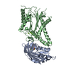
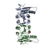
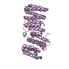
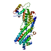
| Title | THE CRYSTAL STRUCTURE OF ZEAMATIN. | ||||||
 Components Components | ZEAMATIN | ||||||
 Keywords Keywords |  ANTIFUNGAL PROTEIN / beta sandwich ANTIFUNGAL PROTEIN / beta sandwich | ||||||
| Function / homology |  Function and homology information: / : / defense response to fungus / defense response / killing of cells of another organism / proteolysis / extracellular region Function and homology information: / : / defense response to fungus / defense response / killing of cells of another organism / proteolysis / extracellular regionSimilarity search - Function | ||||||
| Biological species |  Zea mays (maize) Zea mays (maize) | ||||||
| Method | X-RAY DIFFRACTION / Resolution: 2.5 Å | ||||||
 Authors Authors | Batalia, M.A. / Monzingo, A.F. / Ernst, S. / Roberts, W. / Robertus, J.D. | ||||||
 Citation Citation | Journal: Nat.Struct.Biol. / Year: 1996 Title: The crystal structure of the antifungal protein zeamatin, a member of the thaumatin-like, PR-5 protein family. Authors: Batalia, M.A. / Monzingo, A.F. / Ernst, S. / Roberts, W. / Robertus, J.D. | ||||||
| History |
|
- Structure visualization
| Structure viewer | Molecule: MolmilJmol/JSmol |
|---|
- Downloads & links
-Download
| PDBx/mmCIF format | 1du5.cif.gz | 82.5 KB | Display | PDBx/mmCIF format |
|---|---|---|---|---|
| PDB format | pdb1du5.ent.gz | 66.9 KB | Display | PDB format |
| PDBx/mmJSON format | 1du5.json.gz | Tree view | PDBx/mmJSON format | |
| Others |  Other downloads Other downloads |
-Validation report
| Arichive directory | https://data.pdbj.org/pub/pdb/validation_reports/du/1du5ftp://data.pdbj.org/pub/pdb/validation_reports/du/1du5 | HTTPS FTP |
|---|
-Related structure data
| Similar structure data |
|---|


-Links
 PDBj
PDBj
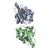
- Assembly
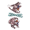
| Deposited unit | 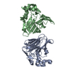
| ||||||||
|---|---|---|---|---|---|---|---|---|---|
| 1 |
| ||||||||
| Unit cell |
|
-Components
| #1: Protein | Mass: 22078.777 Da / Num. of mol.: 2 / Source method: isolated from a natural source / Source: (natural) Zea mays (maize) / References: UniProt: P33679#2: Water | ChemComp-HOH / | Water Mass: 18.015 Da / Num. of mol.: 39 / Source method: isolated from a natural source / Formula: H2O Mass: 18.015 Da / Num. of mol.: 39 / Source method: isolated from a natural source / Formula: H2O |
|---|
-Experimental details
-Experiment
| Experiment | Method: X-RAY DIFFRACTION / Number of used crystals: 1 |
|---|
- Sample preparation
| Crystal | Density Matthews: 2.06 Å3/Da / Density % sol: 40.17 % | |||||||||||||||
|---|---|---|---|---|---|---|---|---|---|---|---|---|---|---|---|---|
| Crystal grow | Temperature: 298 K / Method: vapor diffusion, hanging drop / pH: 6.5 Details: PEG 8000, potassium phosphate, pH 6.5, VAPOR DIFFUSION, HANGING DROP, temperature 298.0K | |||||||||||||||
| Crystal grow | *PLUS Method: vapor diffusion | |||||||||||||||
| Components of the solutions | *PLUS
|
-Data collection
| Diffraction | Mean temperature: 298 K |
|---|---|
| Diffraction source | Source: ROTATING ANODE / Type: ELLIOTT GX-20 / Wavelength: 1.5418 |
| Detector | Type: SDMS / Detector: AREA DETECTOR / Date: May 4, 1993 |
| Radiation | Protocol: SINGLE WAVELENGTH / Monochromatic (M) / Laue (L): M / Scattering type: x-ray |
| Radiation wavelength | Wavelength: 1.5418 Å / Relative weight: 1 |
| Reflection | Resolution: 2.5→20 Å / Num. all: 12577 / Num. obs: 12219 / % possible obs: 97.2 % / Observed criterion σ(I): 0 / Redundancy: 6.4 % / Biso Wilson estimate: 20.2 Å2 / Rmerge(I) obs: 0.085 / Net I/σ(I): 19.8 |
| Reflection shell | Resolution: 2.5→2.69 Å / Redundancy: 4.5 % / Rmerge(I) obs: 0.152 / Num. unique all: 2306 / % possible all: 93.2 |
| Reflection shell | *PLUS % possible obs: 93.2 % |
- Processing
| Software |
| |||||||||||||||||||||||||
|---|---|---|---|---|---|---|---|---|---|---|---|---|---|---|---|---|---|---|---|---|---|---|---|---|---|---|
| Refinement | Resolution: 2.5→5 Å / σ(F): 0 / σ(I): 0 / Stereochemistry target values: Engh & Huber
| |||||||||||||||||||||||||
| Refinement step | Cycle: LAST / Resolution: 2.5→5 Å
| |||||||||||||||||||||||||
| Refine LS restraints |
| |||||||||||||||||||||||||
| Software | *PLUS Name: X-PLOR / Version: 3.1 / Classification: refinement | |||||||||||||||||||||||||
| Refinement | *PLUS | |||||||||||||||||||||||||
| Solvent computation | *PLUS | |||||||||||||||||||||||||
| Displacement parameters | *PLUS | |||||||||||||||||||||||||
| Refine LS restraints | *PLUS
|
