 Movie
Movie Controller

[English] 日本語
 Yorodumi
- PDB-1dt7: SOLUTION STRUCTURE OF THE C-TERMINAL NEGATIVE REGULATORY DOMAIN O... -
+ Open data

- Basic information
| Entry | Database: PDB / ID: 1dt7 | ||||||
|---|---|---|---|---|---|---|---|
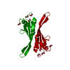
| Title | SOLUTION STRUCTURE OF THE C-TERMINAL NEGATIVE REGULATORY DOMAIN OF P53 IN A COMPLEX WITH CA2+-BOUND S100B(BB) | ||||||
 Components Components |
| ||||||
 Keywords Keywords |  SIGNALING PROTEIN / S100B / p53 / C-terminal domain of p53 / Calcium-binding / EF-hand / S100 protein / four helix bundle / helix loop helix SIGNALING PROTEIN / S100B / p53 / C-terminal domain of p53 / Calcium-binding / EF-hand / S100 protein / four helix bundle / helix loop helix | ||||||
| Function / homology |  Function and homology information Function and homology informationnegative regulation of skeletal muscle cell differentiation / TRAF6 mediated NF-kB activation / Advanced glycosylation endproduct receptor signaling / TAK1-dependent IKK and NF-kappa-B activation / adaptive thermogenesis / sympathetic neuron projection extension / positive regulation of myelination / RAGE receptor binding / response to methylmercury / ion binding ...negative regulation of skeletal muscle cell differentiation / TRAF6 mediated NF-kB activation / Advanced glycosylation endproduct receptor signaling / TAK1-dependent IKK and NF-kappa-B activation / adaptive thermogenesis / sympathetic neuron projection extension / positive regulation of myelination / RAGE receptor binding / response to methylmercury / ion binding / astrocyte differentiation / Loss of function of TP53 in cancer due to loss of tetramerization ability / Regulation of TP53 Expression / : / : / signal transduction by p53 class mediator / negative regulation of G1 to G0 transition / negative regulation of glucose catabolic process to lactate via pyruvate / Transcriptional activation of cell cycle inhibitor p21 / regulation of intrinsic apoptotic signaling pathway by p53 class mediator / Activation of NOXA and translocation to mitochondria / negative regulation of pentose-phosphate shunt / ATP-dependent DNA/DNA annealing activity / negative regulation of helicase activity / S100 protein binding / regulation of cell cycle G2/M phase transition / intrinsic apoptotic signaling pathway in response to hypoxia / regulation of fibroblast apoptotic process / oxidative stress-induced premature senescence / oligodendrocyte apoptotic process / negative regulation of miRNA processing / positive regulation of thymocyte apoptotic process / glucose catabolic process to lactate via pyruvate / regulation of tissue remodeling / positive regulation of mitochondrial membrane permeability / negative regulation of mitophagy / positive regulation of programmed necrotic cell death / mRNA transcription / bone marrow development / circadian behavior / histone deacetylase regulator activity / T cell proliferation involved in immune response / regulation of mitochondrial membrane permeability involved in apoptotic process / RUNX3 regulates CDKN1A transcription / germ cell nucleus / regulation of DNA damage response, signal transduction by p53 class mediator / TP53 regulates transcription of additional cell cycle genes whose exact role in the p53 pathway remain uncertain / TP53 Regulates Transcription of Death Receptors and Ligands / Activation of PUMA and translocation to mitochondria / negative regulation of neuroblast proliferation / DNA damage response, signal transduction by p53 class mediator resulting in transcription of p21 class mediator / neuron projection extension / Formation of Senescence-Associated Heterochromatin Foci (SAHF) / negative regulation of glial cell proliferation / Regulation of TP53 Activity through Association with Co-factors / positive regulation of execution phase of apoptosis / regulation of neuronal synaptic plasticity / mitochondrial DNA repair / T cell lineage commitment / negative regulation of DNA replication / ER overload response / negative regulation of telomerase activity / B cell lineage commitment / thymocyte apoptotic process / positive regulation of cardiac muscle cell apoptotic process / TP53 regulates transcription of several additional cell death genes whose specific roles in p53-dependent apoptosis remain uncertain / TP53 Regulates Transcription of Caspase Activators and Caspases / entrainment of circadian clock by photoperiod / cardiac septum morphogenesis / PI5P Regulates TP53 Acetylation / Association of TriC/CCT with target proteins during biosynthesis / Zygotic genome activation (ZGA) / necroptotic process / positive regulation of release of cytochrome c from mitochondria / rRNA transcription / TP53 Regulates Transcription of Genes Involved in Cytochrome C Release / TFIID-class transcription factor complex binding / mitophagy / SUMOylation of transcription factors / intrinsic apoptotic signaling pathway by p53 class mediator / general transcription initiation factor binding / neuroblast proliferation / cellular response to actinomycin D / Transcriptional Regulation by VENTX / response to X-ray / DNA damage response, signal transduction by p53 class mediator / replicative senescence / intrinsic apoptotic signaling pathway in response to endoplasmic reticulum stress / chromosome organization / gastrulation / cellular response to UV-C / response to inorganic substance / hematopoietic stem cell differentiation / intrinsic apoptotic signaling pathway in response to DNA damage by p53 class mediator / negative regulation of reactive oxygen species metabolic process / positive regulation of RNA polymerase II transcription preinitiation complex assembly / MDM2/MDM4 family protein binding / glial cell proliferation / embryonic organ development / cellular response to glucose starvationSimilarity search - Function | ||||||
| Biological species |  Rattus norvegicus (Norway rat) Rattus norvegicus (Norway rat) | ||||||
| Method | SOLUTION NMR | ||||||
 Authors Authors | Rustandi, R.R. / Baldisseri, D.M. / Weber, D.J. | ||||||
 Citation Citation | Journal: Nat.Struct.Biol. / Year: 2000 Title: Structure of the negative regulatory domain of p53 bound to S100B(betabeta). Authors: Rustandi, R.R. / Baldisseri, D.M. / Weber, D.J. | ||||||
| History |
|
- Structure visualization
| Structure viewer | Molecule: MolmilJmol/JSmol |
|---|
- Downloads & links
-Download
| PDBx/mmCIF format | 1dt7.cif.gz | 2.8 MB | Display | PDBx/mmCIF format |
|---|---|---|---|---|
| PDB format | pdb1dt7.ent.gz | 2.4 MB | Display | PDB format |
| PDBx/mmJSON format | 1dt7.json.gz | Tree view | PDBx/mmJSON format | |
| Others |  Other downloads Other downloads |
-Validation report
| Arichive directory | https://data.pdbj.org/pub/pdb/validation_reports/dt/1dt7ftp://data.pdbj.org/pub/pdb/validation_reports/dt/1dt7 | HTTPS FTP |
|---|
-Related structure data
| Similar structure data | |
|---|---|
| Other databases |
|


-Links
 PDBj
PDBj


- Assembly
| Deposited unit | 
| |||||||||
|---|---|---|---|---|---|---|---|---|---|---|
| 1 |
| |||||||||
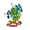
| NMR ensembles |
|
-Components
| #1: Protein | Mass: 10758.048 Da / Num. of mol.: 2 / Fragment: BETA CHAIN Source method: isolated from a genetically manipulated source Source: (gene. exp.) Rattus norvegicus (Norway rat) / Organ: BRAIN / Plasmid: HMS174(DE3) / Production host:  Escherichia coli (E. coli) / References: UniProt: P04631 Escherichia coli (E. coli) / References: UniProt: P04631#2: Protein/peptide | P53Mass: 2596.080 Da / Num. of mol.: 2 / Fragment: C-TERMINAL PEPTIDE / Source method: obtained synthetically Details: THIS PEPTIDE WAS CHEMICALLY SYNTHESIZED. THE SEQUENCE OF THIS PEPTIDE NATURALLY OCCURS IN HUMANS (HOMO SAPIENS). References: UniProt: P04637 #3: Chemical | ChemComp-CA /   Mass: 40.078 Da / Num. of mol.: 4 / Source method: obtained synthetically / Formula: Ca Mass: 40.078 Da / Num. of mol.: 4 / Source method: obtained synthetically / Formula: Ca |
|---|
-Experimental details
-Experiment
| Experiment | Method: SOLUTION NMR | ||||||||||||||||||||||||||||
|---|---|---|---|---|---|---|---|---|---|---|---|---|---|---|---|---|---|---|---|---|---|---|---|---|---|---|---|---|---|
| NMR experiment |
| ||||||||||||||||||||||||||||
| NMR details | Text: This structure was determined using triple-resonance NMR spectroscopy |
- Sample preparation
| Details | Contents: 3-6 mM S100 (subunit concentration), 0.1 mM EDTA, 0.34 mM NaN3, 5 mM DTT, 17 mM NaCl, 10 mM d11-tris-HCl, 7% D2O, 6-13 mM CaCl2, 4.8-10 mM p53 peptide Solvent system: 7% D2O, 93% H2O |
|---|---|
| Sample conditions | Ionic strength: 25 mM / pH: 6.5 / Pressure: ambient / Temperature: 310 K |
| Crystal grow | *PLUS Method: other / Details: NMR |
-NMR measurement
| NMR spectrometer | Type: Bruker DMX / Manufacturer: Bruker / Model: DMX / Field strength: 600 MHz |
|---|
- Processing
| NMR software |
| ||||||||||||||||||||
|---|---|---|---|---|---|---|---|---|---|---|---|---|---|---|---|---|---|---|---|---|---|
| NMR representative | Selection criteria: lowest energy | ||||||||||||||||||||
| NMR ensemble | Conformer selection criteria: structure with the lowest energy and the least restraint violations Conformers calculated total number: 40 / Conformers submitted total number: 40 |
