 Movie
Movie Controller

[English] 日本語
 Yorodumi
- PDB-1dqj: CRYSTAL STRUCTURE OF THE ANTI-LYSOZYME ANTIBODY HYHEL-63 COMPLEXE... -
+ Open data

- Basic information
| Entry | Database: PDB / ID: 1dqj | ||||||
|---|---|---|---|---|---|---|---|
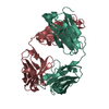
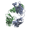
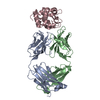
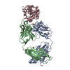
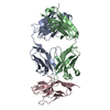
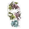
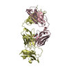
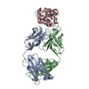
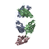
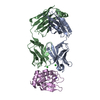
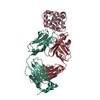
| Title | CRYSTAL STRUCTURE OF THE ANTI-LYSOZYME ANTIBODY HYHEL-63 COMPLEXED WITH HEN EGG WHITE LYSOZYME | ||||||
 Components Components |
| ||||||
 Keywords Keywords |  immune system/hydrolase / antibody / protein-protein complex / hen egg white lysozyme / immune system-hydrolase COMPLEX immune system/hydrolase / antibody / protein-protein complex / hen egg white lysozyme / immune system-hydrolase COMPLEX | ||||||
| Function / homology |  Function and homology information Function and homology informationpositive regulation of B cell activation / early endosome to late endosome transport / humoral immune response mediated by circulating immunoglobulin / phagocytosis, recognition / positive regulation of type IIa hypersensitivity / regulation of proteolysis / positive regulation of type I hypersensitivity / antibody-dependent cellular cytotoxicity / Fc-gamma receptor I complex binding / phagocytosis, engulfment ...positive regulation of B cell activation / early endosome to late endosome transport / humoral immune response mediated by circulating immunoglobulin / phagocytosis, recognition / positive regulation of type IIa hypersensitivity / regulation of proteolysis / positive regulation of type I hypersensitivity / antibody-dependent cellular cytotoxicity / Fc-gamma receptor I complex binding / phagocytosis, engulfment / endosome to lysosome transport / positive regulation of endocytosis / immunoglobulin complex, circulating / IgG immunoglobulin complex / immunoglobulin receptor binding / immunoglobulin mediated immune response / antigen processing and presentation / positive regulation of phagocytosis / complement activation, classical pathway / antigen binding / multivesicular body / B cell differentiation / Antimicrobial peptides / Neutrophil degranulation / beta-N-acetylglucosaminidase activity / response to bacterium / positive regulation of immune response / cell wall macromolecule catabolic process / lysozyme / lysozyme activity / antibacterial humoral response / killing of cells of another organism / defense response to Gram-negative bacterium / defense response to Gram-positive bacterium / defense response to bacterium / endoplasmic reticulum / extracellular space / extracellular region / identical protein binding / plasma membrane / cytosol / cytoplasmSimilarity search - Function | ||||||
| Biological species |  Mus musculus (house mouse) Mus musculus (house mouse) Gallus gallus (chicken) Gallus gallus (chicken) | ||||||
| Method | X-RAY DIFFRACTION / Resolution: 2 Å | ||||||
 Authors Authors | Li, H. / Mariuzza, R.A. | ||||||
 Citation Citation | Journal: Biochemistry / Year: 2000 Title: Three-dimensional structures of the free and antigen-bound Fab from monoclonal antilysozyme antibody HyHEL-63(,). Authors: Li, Y. / Li, H. / Smith-Gill, S.J. / Mariuzza, R.A. | ||||||
| History |
|
- Structure visualization
| Structure viewer | Molecule: MolmilJmol/JSmol |
|---|
- Downloads & links
-Download
| PDBx/mmCIF format | 1dqj.cif.gz | 128.4 KB | Display | PDBx/mmCIF format |
|---|---|---|---|---|
| PDB format | pdb1dqj.ent.gz | 102 KB | Display | PDB format |
| PDBx/mmJSON format | 1dqj.json.gz | Tree view | PDBx/mmJSON format | |
| Others |  Other downloads Other downloads |
-Validation report
| Arichive directory | https://data.pdbj.org/pub/pdb/validation_reports/dq/1dqjftp://data.pdbj.org/pub/pdb/validation_reports/dq/1dqj | HTTPS FTP |
|---|
-Related structure data


-Links
 PDBj
PDBj


- Assembly
| Deposited unit | 
| ||||||||||||
|---|---|---|---|---|---|---|---|---|---|---|---|---|---|
| 1 |
| ||||||||||||
| Unit cell |
| ||||||||||||
| Components on special symmetry positions |
| ||||||||||||
| Details | The biological assembly is a heterodimer with heavy and light chains |
-Components
| #1: Antibody | Mass: 23583.869 Da / Num. of mol.: 1 / Fragment: FAB FRAGMENT Source method: isolated from a genetically manipulated source Source: (gene. exp.) Mus musculus (house mouse) / Plasmid: PET22B / Production host:  Escherichia coli (E. coli) / References: GenBank: 2950241, UniProt: P01837*PLUS Escherichia coli (E. coli) / References: GenBank: 2950241, UniProt: P01837*PLUS |
|---|---|
| #2: Antibody | Mass: 22556.023 Da / Num. of mol.: 1 / Fragment: FAB FRAGMENT Source method: isolated from a genetically manipulated source Source: (gene. exp.) Mus musculus (house mouse) / Plasmid: PET22B / Production host: Escherichia coli (E. coli) / References: UniProt: P01865*PLUS |
| #3: Protein | Mass: 14331.160 Da / Num. of mol.: 1 Source method: isolated from a genetically manipulated source Source: (gene. exp.) Gallus gallus (chicken) / Cell: EGG / Cellular location: CYTOPLASM / Plasmid: PPIC9 / Production host:  Saccharomyces cerevisiae (brewer's yeast) / References: UniProt: P00698, lysozyme Saccharomyces cerevisiae (brewer's yeast) / References: UniProt: P00698, lysozyme |
| #4: Water | ChemComp-HOH / Water Mass: 18.015 Da / Num. of mol.: 467 / Source method: isolated from a natural source / Formula: H2O Mass: 18.015 Da / Num. of mol.: 467 / Source method: isolated from a natural source / Formula: H2O |
-Experimental details
-Experiment
| Experiment | Method: X-RAY DIFFRACTION / Number of used crystals: 1 |
|---|
- Sample preparation
| Crystal | Density Matthews: 2.54 Å3/Da / Density % sol: 51.65 % | |||||||||||||||||||||||||
|---|---|---|---|---|---|---|---|---|---|---|---|---|---|---|---|---|---|---|---|---|---|---|---|---|---|---|
| Crystal grow | Temperature: 298 K / Method: vapor diffusion, hanging drop / pH: 4.6 Details: PEG 4K, Ammonium acetate, sodium acetate, pH 4.6, VAPOR DIFFUSION, HANGING DROP, temperature 298K | |||||||||||||||||||||||||
| Crystal grow | *PLUS | |||||||||||||||||||||||||
| Components of the solutions | *PLUS
|
-Data collection
| Diffraction | Mean temperature: 298 K |
|---|---|
| Diffraction source | Source: ROTATING ANODE / Type: RIGAKU RU200 / Wavelength: 1.5418 |
| Detector | Type: MACSCIENCE / Detector: IMAGE PLATE / Date: May 5, 1998 |
| Radiation | Protocol: SINGLE WAVELENGTH / Monochromatic (M) / Laue (L): M / Scattering type: x-ray |
| Radiation wavelength | Wavelength: 1.5418 Å / Relative weight: 1 |
| Reflection | Resolution: 2→100 Å / Num. obs: 45762 / % possible obs: 92.7 % / Observed criterion σ(F): 2 / Observed criterion σ(I): 2 / Redundancy: 8 % / Biso Wilson estimate: 14.4 Å2 / Rmerge(I) obs: 0.089 / Net I/σ(I): 19.6 |
| Reflection shell | Resolution: 2→2.1 Å / Redundancy: 4 % / Rmerge(I) obs: 0.371 / % possible all: 85 |
| Reflection | *PLUS Num. obs: 45762 / Num. measured all: 366760 |
| Reflection shell | *PLUS % possible obs: 85 % / Mean I/σ(I) obs: 5.1 |
- Processing
| Software |
| |||||||||||||||||||||||||
|---|---|---|---|---|---|---|---|---|---|---|---|---|---|---|---|---|---|---|---|---|---|---|---|---|---|---|
| Refinement | Resolution: 2→100 Å / σ(F): 0 / σ(I): 0 / Stereochemistry target values: Engh & Huber
| |||||||||||||||||||||||||
| Refinement step | Cycle: LAST / Resolution: 2→100 Å
| |||||||||||||||||||||||||
| Refine LS restraints |
| |||||||||||||||||||||||||
| Software | *PLUS Name: 'CNS' / Classification: refinement | |||||||||||||||||||||||||
| Refinement | *PLUS Rfactor Rfree: 0.249 | |||||||||||||||||||||||||
| Solvent computation | *PLUS | |||||||||||||||||||||||||
| Displacement parameters | *PLUS Biso mean: 25.5 Å2 | |||||||||||||||||||||||||
| Refine LS restraints | *PLUS
|
