- PDB-1dpu: SOLUTION STRUCTURE OF THE C-TERMINAL DOMAIN OF HUMAN RPA32 COMPLE... -
+
Open data
ID or keywords:
Loading...
-
Basic information
Entry
Database: PDB / ID: 1dpu
Title
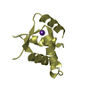
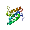
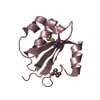
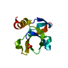
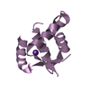
SOLUTION STRUCTURE OF THE C-TERMINAL DOMAIN OF HUMAN RPA32 COMPLEXED WITH UNG2(73-88)
Components
REPLICATION PROTEIN A (RPA32) C-TERMINAL DOMAINDNA replication
URACIL DNA GLYCOSYLASE (UNG2)
Keywords
DNA BINDING PROTEIN / PROTEIN-PEPTIDE COMPLEX / DNA REPAIR
Function / homology
Function and homology information
protein localization to chromosome / DNA replication factor A complex / base-excision repair, AP site formation via deaminated base removal / uracil-DNA glycosylase / depyrimidination / Displacement of DNA glycosylase by APEX1 / Removal of the Flap Intermediate / G-rich strand telomeric DNA binding / regulation of DNA damage checkpoint / Mismatch repair (MMR) directed by MSH2:MSH3 (MutSbeta) ...protein localization to chromosome / DNA replication factor A complex / base-excision repair, AP site formation via deaminated base removal / uracil-DNA glycosylase / depyrimidination / Displacement of DNA glycosylase by APEX1 / Removal of the Flap Intermediate / G-rich strand telomeric DNA binding / regulation of DNA damage checkpoint / Mismatch repair (MMR) directed by MSH2:MSH3 (MutSbeta) / Mismatch repair (MMR) directed by MSH2:MSH6 (MutSalpha) / isotype switching / Removal of the Flap Intermediate from the C-strand / uracil DNA N-glycosylase activity / HDR through Single Strand Annealing (SSA) / Impaired BRCA2 binding to RAD51 / regulation of double-strand break repair via homologous recombination / telomeric DNA binding / Presynaptic phase of homologous DNA pairing and strand exchange / ribosomal small subunit binding / PCNA-Dependent Long Patch Base Excision Repair / mismatch repair / Activation of the pre-replicative complex / Regulation of HSF1-mediated heat shock response / HSF1 activation / somatic hypermutation of immunoglobulin genes / Activation of ATR in response to replication stress / Recognition and association of DNA glycosylase with site containing an affected pyrimidine / Cleavage of the damaged pyrimidine / mitotic G1 DNA damage checkpoint signaling / telomere maintenance / Translesion synthesis by REV1 / Translesion synthesis by POLK / Translesion synthesis by POLI / Gap-filling DNA repair synthesis and ligation in GG-NER / Chromatin modifications during the maternal to zygotic transition (MZT) / Recognition of DNA damage by PCNA-containing replication complex / nucleotide-excision repair / Fanconi Anemia Pathway / Termination of translesion DNA synthesis / Translesion Synthesis by POLH / double-strand break repair via homologous recombination / base-excision repair / HDR through Homologous Recombination (HRR) / G2/M DNA damage checkpoint / Dual Incision in GG-NER / PML body / Meiotic recombination / Formation of Incision Complex in GG-NER / Dual incision in TC-NER / Gap-filling DNA repair synthesis and ligation in TC-NER / single-stranded DNA binding / Processing of DNA double-strand break ends / protein phosphatase binding / Regulation of TP53 Activity through Phosphorylation / DNA replication / damaged DNA binding / chromosome, telomeric region / nuclear body / ubiquitin protein ligase binding / chromatin / negative regulation of apoptotic process / enzyme binding / mitochondrion / nucleoplasm / nucleus Similarity search - Function
Replication factor A protein 2 / Replication protein A, C-terminal / Replication protein A C terminal / Replication factor A protein-like / Uracil-DNA glycosylase family 1 / Uracil-DNA glycosylase, active site / Uracil-DNA glycosylase signature. / UreE urease accessory protein, C-terminal domain / Uracil DNA glycosylase superfamily / Uracil-DNA glycosylase-like ...Replication factor A protein 2 / Replication protein A, C-terminal / Replication protein A C terminal / Replication factor A protein-like / Uracil-DNA glycosylase family 1 / Uracil-DNA glycosylase, active site / Uracil-DNA glycosylase signature. / UreE urease accessory protein, C-terminal domain / Uracil DNA glycosylase superfamily / Uracil-DNA glycosylase-like / Uracil DNA glycosylase superfamily / Uracil-DNA glycosylase-like domain superfamily / Winged helix-like DNA-binding domain superfamily/Winged helix DNA-binding domain / Arc Repressor Mutant, subunit A / Winged helix DNA-binding domain superfamily / Winged helix-like DNA-binding domain superfamily / Nucleic acid-binding, OB-fold / Orthogonal Bundle / Mainly Alpha Similarity search - Domain/homology
REPLICATIONPROTEINA (RPA32) C-TERMINALDOMAIN / DNA replication
Mass: 10585.656 Da / Num. of mol.: 1 / Fragment: C-TERMINAL DOMAIN (RESIDUES 172-270) Source method: isolated from a genetically manipulated source Source: (gene. exp.) Homo sapiens (human) / Plasmid: PET15B / Production host: Escherichia coli (E. coli) / References: UniProt: P15927
#2: Protein/peptide
URACILDNAGLYCOSYLASE (UNG2)
Mass: 1826.222 Da / Num. of mol.: 1 / Fragment: RESIDUES 73-88 / Source method: obtained synthetically Details: THIS PEPTIDE SEQUENCE IS FOUND IN THE NUCLEAR [UNG2(73-88)] AND MITOCHONDRIAL [UNG1(64-79)] FORMS OF HUMAN URACIL-DNA GLYCOSYLASE References: UniProt: P13051
-
Experimental details
-
Experiment
Experiment
Method: SOLUTION NMR
NMR experiment
Conditions-ID
Experiment-ID
Solution-ID
Type
1
1
1
2D NOESY
1
2
1
3D 13C- SEPARATED NOESY
1
3
1
3D 15N- SEPARATED NOESY
1
4
1
DQF- COSY
1
5
1
HNHA
1
6
1
HNHB
1
7
1
HACAHB -COSY
1
8
1
FILTER-EDITED NOESY
1
9
1
DOUBLE-HALF FILTERED NOESY
NMR details
Text: THE STRUCTURE WAS DETERMINED USING DOUBLE- AND TRIPLE-RESONANCE NMR SPECTROSCOPY ***
-
Sample preparation
Details
Contents: 1MM RPA32(172-270) U- 15N,13C; 1.2 MM UNG2(73- 88) NA; 25MM PHOSPHATE BUFFER NA; 50MM NACL; 5 MM DTT NA
Conformer selection criteria: structures with the least restraint violations Conformers calculated total number: 50 / Conformers submitted total number: 30
+
About Yorodumi
-
News
-
Feb 9, 2022. New format data for meta-information of EMDB entries
New format data for meta-information of EMDB entries
Version 3 of the EMDB header file is now the official format.
The previous official version 1.9 will be removed from the archive.
In the structure databanks used in Yorodumi, some data are registered as the other names, "COVID-19 virus" and "2019-nCoV". Here are the details of the virus and the list of structure data.
Jan 31, 2019. EMDB accession codes are about to change! (news from PDBe EMDB page)
EMDB accession codes are about to change! (news from PDBe EMDB page)
The allocation of 4 digits for EMDB accession codes will soon come to an end. Whilst these codes will remain in use, new EMDB accession codes will include an additional digit and will expand incrementally as the available range of codes is exhausted. The current 4-digit format prefixed with “EMD-” (i.e. EMD-XXXX) will advance to a 5-digit format (i.e. EMD-XXXXX), and so on. It is currently estimated that the 4-digit codes will be depleted around Spring 2019, at which point the 5-digit format will come into force.
The EM Navigator/Yorodumi systems omit the EMD- prefix.
Related info.:Q: What is EMD? / ID/Accession-code notation in Yorodumi/EM Navigator
Yorodumi is a browser for structure data from EMDB, PDB, SASBDB, etc.
This page is also the successor to EM Navigator detail page, and also detail information page/front-end page for Omokage search.
The word "yorodu" (or yorozu) is an old Japanese word meaning "ten thousand". "mi" (miru) is to see.
Related info.:EMDB / PDB / SASBDB / Comparison of 3 databanks / Yorodumi Search / Aug 31, 2016. New EM Navigator & Yorodumi / Yorodumi Papers / Jmol/JSmol / Function and homology information / Changes in new EM Navigator and Yorodumi
 Movie
Movie Controller

 Yorodumi
 Open data
 Basic information
 Components
 Keywords
 DNA BINDING PROTEIN / PROTEIN-PEPTIDE COMPLEX /
  Function and homology information
 Authors
 Citation
 Structure visualization
 Downloads & links
 Other downloads


 PDBj
PDBj


 Assembly
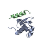

 Sample preparation
 Processing