 Movie
Movie Controller

[English] 日本語
 Yorodumi
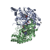
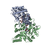
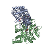
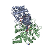
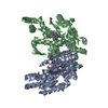
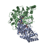
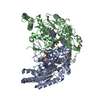
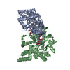
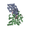
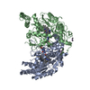
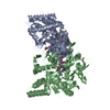
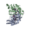
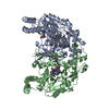
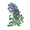
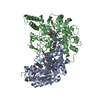
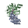
- PDB-1dm7: BOVINE ENDOTHELIAL NITRIC OXIDE SYNTHASE HEME DOMAIN COMPLEXED WI... -
+ Open data

- Basic information
| Entry | Database: PDB / ID: 1dm7 | ||||||
|---|---|---|---|---|---|---|---|
| Title | BOVINE ENDOTHELIAL NITRIC OXIDE SYNTHASE HEME DOMAIN COMPLEXED WITH HOMOARGININE (H4B FREE) | ||||||
 Components Components | NITRIC OXIDE SYNTHASE | ||||||
 Keywords Keywords | OXIDOREDUCTASE / ALPHA-BETA FOLD | ||||||
| Function / homology |  Function and homology information Function and homology informationcellular response to laminar fluid shear stress / positive regulation of guanylate cyclase activity / negative regulation of leukocyte cell-cell adhesion / nitric-oxide synthase (NADPH) / nitric-oxide synthase activity / nitric oxide mediated signal transduction / arginine catabolic process / negative regulation of extrinsic apoptotic signaling pathway via death domain receptors / mitochondrion organization / negative regulation of blood pressure ...cellular response to laminar fluid shear stress / positive regulation of guanylate cyclase activity / negative regulation of leukocyte cell-cell adhesion / nitric-oxide synthase (NADPH) / nitric-oxide synthase activity / nitric oxide mediated signal transduction / arginine catabolic process / negative regulation of extrinsic apoptotic signaling pathway via death domain receptors / mitochondrion organization / negative regulation of blood pressure / nitric oxide biosynthetic process / response to hormone / caveola / blood coagulation / FMN binding / flavin adenine dinucleotide binding / NADP binding / response to lipopolysaccharide / cytoskeleton / calmodulin binding / heme binding / Golgi apparatus / metal ion binding / nucleus / plasma membrane / cytosolSimilarity search - Function | ||||||
| Biological species |  Bos taurus (cattle) Bos taurus (cattle) | ||||||
| Method | X-RAY DIFFRACTION / SYNCHROTRON / Resolution: 2.1 Å | ||||||
 Authors Authors | Raman, C.S. / Li, H. / Martasek, P. / Southan, G.J. / Masters, B.S.S. / Poulos, T.L. | ||||||
 Citation Citation | Journal: To be Published Title: Crystal Structures of Bovine Endothelial Nitric Oxide Synthase Heme Domain Complexed With Various Inhibitors Authors: Raman, C.S. / Li, H. / Martasek, P. / Southan, G.J. / Masters, B.S.S. / Poulos, T.L. #1: Journal: Cell(Cambridge,Mass.) / Year: 1998Title: Crystal Structure of Constitutive Endothelial Nitric Oxide Synthase: A Paradigm for Pterin Function Involving a Novel Metal Center Authors: Raman, C.S. / Li, H. / Martasek, P. / Kral, V. / Masters, B.S.S. / Poulos, T.L. #2: Journal: Science / Year: 1998Title: Structure of Nitric Oxide Synthase Oxygenase Dimer with Pterin and Substrate Authors: Crane, B.R. / Arvai, A.S. / Ghosh, D.K. / Wu, C. / Getzoff, E.D. / Stuehr, D.J. / Tainer, A. #3: Journal: Biochemistry / Year: 1999Title: Efficient Formation of Nitric Oxide from Selective Oxidation of N-Aryl N'- Hydroxyguanidines by Inducible Nitric Oxide Synthase Authors: Renodon-Corniere, A. / Boucher, J.-L. / Dijols, S. / Stuehr, D.J. / Mansuy, D. | ||||||
| History |
|
- Structure visualization
| Structure viewer | Molecule: MolmilJmol/JSmol |
|---|
- Downloads & links
-Download
| PDBx/mmCIF format | 1dm7.cif.gz | 192.8 KB | Display | PDBx/mmCIF format |
|---|---|---|---|---|
| PDB format | pdb1dm7.ent.gz | 150.3 KB | Display | PDB format |
| PDBx/mmJSON format | 1dm7.json.gz | Tree view | PDBx/mmJSON format | |
| Others |  Other downloads Other downloads |
-Validation report
| Arichive directory | https://data.pdbj.org/pub/pdb/validation_reports/dm/1dm7ftp://data.pdbj.org/pub/pdb/validation_reports/dm/1dm7 | HTTPS FTP |
|---|
-Related structure data


-Links
 PDBj
PDBj


- Assembly
| Deposited unit | 
| ||||||||
|---|---|---|---|---|---|---|---|---|---|
| 1 |
| ||||||||
| Unit cell |
|
-Components
-Protein , 1 types, 2 molecules AB
| #1: Protein | Mass: 49710.105 Da / Num. of mol.: 2 / Fragment: HEME DOMAIN Source method: isolated from a genetically manipulated source Source: (gene. exp.) Bos taurus (cattle) / Cell: ENDOTHELIAL / Production host:  Escherichia coli (E. coli) / References: UniProt: P29473, nitric-oxide synthase (NADPH) Escherichia coli (E. coli) / References: UniProt: P29473, nitric-oxide synthase (NADPH) |
|---|
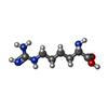
-Non-polymers , 7 types, 423 molecules 


| #2: Chemical | Acetate Mass: 59.044 Da / Num. of mol.: 2 / Source method: obtained synthetically / Formula: C2H3O2 Mass: 59.044 Da / Num. of mol.: 2 / Source method: obtained synthetically / Formula: C2H3O2#3: Chemical | Cacodylic acid Mass: 136.989 Da / Num. of mol.: 2 / Source method: obtained synthetically / Formula: C2H6AsO2 Mass: 136.989 Da / Num. of mol.: 2 / Source method: obtained synthetically / Formula: C2H6AsO2#4: Chemical | Heme B Mass: 616.487 Da / Num. of mol.: 2 / Source method: obtained synthetically / Formula: C34H32FeN4O4 Mass: 616.487 Da / Num. of mol.: 2 / Source method: obtained synthetically / Formula: C34H32FeN4O4#5: Chemical | Homoarginine Type: L-peptide linking / Mass: 188.228 Da / Num. of mol.: 2 / Source method: obtained synthetically / Formula: C7H16N4O2 Type: L-peptide linking / Mass: 188.228 Da / Num. of mol.: 2 / Source method: obtained synthetically / Formula: C7H16N4O2#6: Chemical | ChemComp-GOL / Glycerol Mass: 92.094 Da / Num. of mol.: 5 / Source method: obtained synthetically / Formula: C3H8O3 Mass: 92.094 Da / Num. of mol.: 5 / Source method: obtained synthetically / Formula: C3H8O3#7: Chemical | ChemComp-ZN / |  Mass: 65.409 Da / Num. of mol.: 1 / Source method: obtained synthetically / Formula: Zn Mass: 65.409 Da / Num. of mol.: 1 / Source method: obtained synthetically / Formula: Zn#8: Water | ChemComp-HOH / | WaterMass: 18.015 Da / Num. of mol.: 409 / Source method: isolated from a natural source / Formula: H2O |
|---|
-Details
| Nonpolymer details | ZN ION SITS RIGHT ON THE NCS 2-FOLD THAT DEFINES THE DIMER INTERFACE AND IS COORDINATED ...ZN ION SITS RIGHT ON THE NCS 2-FOLD THAT DEFINES THE DIMER INTERFACE AND IS COORDINATE |
|---|
-Experimental details
-Experiment
| Experiment | Method: X-RAY DIFFRACTION / Number of used crystals: 1 |
|---|
- Sample preparation
| Crystal | Density Matthews: 2.46 Å3/Da / Density % sol: 49.97 % |
|---|---|
| Crystal grow | Temperature: 280 K / Method: vapor diffusion, sitting drop / pH: 6.5 Details: PEG 4000, CACODYLATE, MAGNESIUM ACETATE, pH 6.5, VAPOR DIFFUSION, SITTING DROP, temperature 280K |
-Data collection
| Diffraction | Mean temperature: 100 K |
|---|---|
| Diffraction source | Source: SYNCHROTRON / Site: SSRL  / Beamline: BL7-1 / Wavelength: 1.08 / Beamline: BL7-1 / Wavelength: 1.08 |
| Detector | Type: MARRESEARCH / Detector: IMAGE PLATE / Date: Jan 1, 1999 |
| Radiation | Protocol: SINGLE WAVELENGTH / Monochromatic (M) / Laue (L): M / Scattering type: x-ray |
| Radiation wavelength | Wavelength: 1.08 Å / Relative weight: 1 |
| Reflection | Resolution: 2→30 Å / Num. obs: 66766 / % possible obs: 98.8 % / Observed criterion σ(I): -3 / Redundancy: 3.2 % / Biso Wilson estimate: 36.4 Å2 / Rmerge(I) obs: 0.064 / Net I/σ(I): 7.1 |
| Reflection shell | Resolution: 2.1→2.24 Å / Redundancy: 3.2 % / Rmerge(I) obs: 0.47 / % possible all: 96.7 |
- Processing
| Software |
| ||||||||||||||||||||||||||||||||||||||||||||||||||||||||||||
|---|---|---|---|---|---|---|---|---|---|---|---|---|---|---|---|---|---|---|---|---|---|---|---|---|---|---|---|---|---|---|---|---|---|---|---|---|---|---|---|---|---|---|---|---|---|---|---|---|---|---|---|---|---|---|---|---|---|---|---|---|---|
| Refinement | Resolution: 2.1→28.4 Å / Rfactor Rfree error: 0.005 / Data cutoff high absF: 2579569.55 / Data cutoff low absF: 0 / Isotropic thermal model: RESTRAINED / Cross valid method: THROUGHOUT / σ(F): 0 / Stereochemistry target values: ENGH & HUBER Details: RESIDUES 39 TO 66 OF MOLECULE A AND RESIDUES 39 TO 68 IN MOLECULE B ARE NOT VISIBLE IN THE ELECTRON DENSITY. RESIDUES 108-120 ARE DISORDERED, BUT THE TENTATIVE MODEL WAS INCLUDED IN THE REFINEMENT.
| ||||||||||||||||||||||||||||||||||||||||||||||||||||||||||||
| Solvent computation | Solvent model: FLAT MODEL / Bsol: 54.8 Å2 / ksol: 0.363 e/Å3 | ||||||||||||||||||||||||||||||||||||||||||||||||||||||||||||
| Displacement parameters | Biso mean: 51.4 Å2
| ||||||||||||||||||||||||||||||||||||||||||||||||||||||||||||
| Refine analyze |
| ||||||||||||||||||||||||||||||||||||||||||||||||||||||||||||
| Refinement step | Cycle: LAST / Resolution: 2.1→28.4 Å
| ||||||||||||||||||||||||||||||||||||||||||||||||||||||||||||
| Refine LS restraints |
| ||||||||||||||||||||||||||||||||||||||||||||||||||||||||||||
| LS refinement shell | Resolution: 2.1→2.18 Å / Rfactor Rfree error: 0.025 / Total num. of bins used: 10
|
