 Movie
Movie Controller


+ Open data

- Basic information
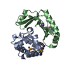
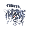
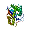
| Entry | Database: PDB / ID: 1dix | ||||||
|---|---|---|---|---|---|---|---|
| Title | CRYSTAL STRUCTURE OF RNASE LE | ||||||
 Components Components | EXTRACELLULAR RIBONUCLEASE LE | ||||||
 Keywords Keywords |  HYDROLASE / ALPHA PLUS BETA HYDROLASE / ALPHA PLUS BETA | ||||||
| Function / homology |  Function and homology informationribonuclease T2 / ribonuclease T2 activity / cell wall / RNA catabolic process / RNA endonuclease activity / lyase activity / extracellular space / RNA binding / extracellular region Function and homology informationribonuclease T2 / ribonuclease T2 activity / cell wall / RNA catabolic process / RNA endonuclease activity / lyase activity / extracellular space / RNA binding / extracellular regionSimilarity search - Function | ||||||
| Biological species |  Solanum lycopersicum (tomato) Solanum lycopersicum (tomato) | ||||||
| Method | X-RAY DIFFRACTION / MIR / Resolution: 1.65 Å | ||||||
 Authors Authors | Tanaka, N. / Nakamura, K.T. | ||||||
 Citation Citation | Journal: J.Mol.Biol. / Year: 2000 Title: Crystal structure of a plant ribonuclease, RNase LE. Authors: Tanaka, N. / Arai, J. / Inokuchi, N. / Koyama, T. / Ohgi, K. / Irie, M. / Nakamura, K.T. #1: Journal: Protein Pept.Lett. / Year: 1999Title: Crystallization and Preliminary X-ray Crystallographic Studies of Ribonuclease LE from Lycopersicon esculentum Authors: Tanaka, N. / Arai, J. / Inokuchi, N. / Koyama, T. / Ohgi, K. / Irie, M. / Nakamura, K.T. | ||||||
| History |
|
- Structure visualization
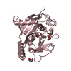
| Structure viewer | Molecule: MolmilJmol/JSmol |
|---|
- Downloads & links
-Download
| PDBx/mmCIF format | 1dix.cif.gz | 51.8 KB | Display | PDBx/mmCIF format |
|---|---|---|---|---|
| PDB format | pdb1dix.ent.gz | 39.7 KB | Display | PDB format |
| PDBx/mmJSON format | 1dix.json.gz | Tree view | PDBx/mmJSON format | |
| Others |  Other downloads Other downloads |
-Validation report
| Arichive directory | https://data.pdbj.org/pub/pdb/validation_reports/di/1dixftp://data.pdbj.org/pub/pdb/validation_reports/di/1dix | HTTPS FTP |
|---|
-Related structure data
| Similar structure data |
|---|


-Links
 PDBj
PDBj- Assembly
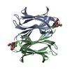
| Deposited unit | 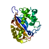
| ||||||||
|---|---|---|---|---|---|---|---|---|---|
| 1 |
| ||||||||
| Unit cell |
|
-Components
| #1: Protein | Mass: 22912.193 Da / Num. of mol.: 1 Source method: isolated from a genetically manipulated source Source: (gene. exp.) Solanum lycopersicum (tomato) / Production host:  Saccharomyces cerevisiae (brewer's yeast) / References: UniProt: P80022, EC: 3.1.27.1 Saccharomyces cerevisiae (brewer's yeast) / References: UniProt: P80022, EC: 3.1.27.1 |
|---|---|
| #2: Water | ChemComp-HOH / Water Mass: 18.015 Da / Num. of mol.: 136 / Source method: isolated from a natural source / Formula: H2O Mass: 18.015 Da / Num. of mol.: 136 / Source method: isolated from a natural source / Formula: H2O |
-Experimental details
-Experiment
| Experiment | Method: X-RAY DIFFRACTION / Number of used crystals: 2 |
|---|
- Sample preparation
| Crystal | Density Matthews: 2.09 Å3/Da / Density % sol: 41.27 % | |||||||||||||||||||||||||
|---|---|---|---|---|---|---|---|---|---|---|---|---|---|---|---|---|---|---|---|---|---|---|---|---|---|---|
| Crystal grow | Temperature: 293 K / Method: vapor diffusion, hanging drop / pH: 5 Details: PEG1540, CITRATE, pH 5.0, VAPOR DIFFUSION, HANGING DROP, temperature 293K | |||||||||||||||||||||||||
| Crystal | *PLUS Density % sol: 41 % | |||||||||||||||||||||||||
| Crystal grow | *PLUS | |||||||||||||||||||||||||
| Components of the solutions | *PLUS
|
-Data collection
| Diffraction | Mean temperature: 293 K |
|---|---|
| Diffraction source | Source: ROTATING ANODE / Type: RIGAKU RU200 / Wavelength: 1.5418 |
| Detector | Type: RIGAKU RAXIS IIC / Detector: IMAGE PLATE / Date: Jul 31, 1998 |
| Radiation | Protocol: SINGLE WAVELENGTH / Monochromatic (M) / Laue (L): M / Scattering type: x-ray |
| Radiation wavelength | Wavelength: 1.5418 Å / Relative weight: 1 |
| Reflection | Resolution: 1.65→100 Å / Num. all: 144121 / Num. obs: 22385 / % possible obs: 93.4 % / Redundancy: 6.4 % / Biso Wilson estimate: 26 Å2 / Rmerge(I) obs: 0.049 |
| Reflection | *PLUS Num. measured all: 144121 / Rmerge(I) obs: 0.054 |
- Processing
| Software |
| ||||||||||||||||
|---|---|---|---|---|---|---|---|---|---|---|---|---|---|---|---|---|---|
| Refinement | Method to determine structure: MIR / Resolution: 1.65→40 Å / σ(F): 1 /
| ||||||||||||||||
| Refinement step | Cycle: LAST / Resolution: 1.65→40 Å
| ||||||||||||||||
| Refine LS restraints |
| ||||||||||||||||
| Software | *PLUS Name: REFMAC / Classification: refinement | ||||||||||||||||
| Refinement | *PLUS σ(F): 1 / Num. reflection Rfree: 2296 / % reflection Rfree: 10 % / Rfactor obs: 0.219 | ||||||||||||||||
| Solvent computation | *PLUS | ||||||||||||||||
| Displacement parameters | *PLUS |
