 Movie
Movie Controller

[English] 日本語
 Yorodumi
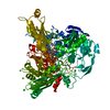
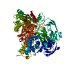
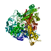
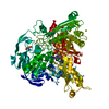
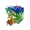
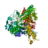
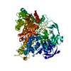
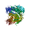
- PDB-1dgj: CRYSTAL STRUCTURE OF THE ALDEHYDE OXIDOREDUCTASE FROM DESULFOVIBR... -
+ Open data

- Basic information
| Entry | Database: PDB / ID: 1dgj | ||||||
|---|---|---|---|---|---|---|---|
| Title | CRYSTAL STRUCTURE OF THE ALDEHYDE OXIDOREDUCTASE FROM DESULFOVIBRIO DESULFURICANS ATCC 27774 | ||||||
 Components Components | ALDEHYDE OXIDOREDUCTASE | ||||||
 Keywords Keywords |  OXIDOREDUCTASE / BETA HALF-BARREL / FOUR-HELIX BUNDLE / BETA BARREL OXIDOREDUCTASE / BETA HALF-BARREL / FOUR-HELIX BUNDLE / BETA BARREL | ||||||
| Function / homology |  Function and homology information Function and homology information2 iron, 2 sulfur cluster binding / oxidoreductase activity / metal ion bindingSimilarity search - Function | ||||||
| Biological species |  Desulfovibrio desulfuricans (bacteria) Desulfovibrio desulfuricans (bacteria) | ||||||
| Method | X-RAY DIFFRACTION / Resolution: 2.8 Å | ||||||
 Authors Authors | Rebelo, J.M. / Macieira, S. / Dias, J.M. / Huber, R. / Romao, M.J. | ||||||
 Citation Citation | Journal: J.Mol.Biol. / Year: 2000 Title: Gene sequence and crystal structure of the aldehyde oxidoreductase from Desulfovibrio desulfuricans ATCC 27774. Authors: Rebelo, J. / Macieira, S. / Dias, J.M. / Huber, R. / Ascenso, C.S. / Rusnak, F. / Moura, J.J. / Moura, I. / Romao, M.J. | ||||||
| History |
|
- Structure visualization
| Structure viewer | Molecule: MolmilJmol/JSmol |
|---|
- Downloads & links
-Download
| PDBx/mmCIF format | 1dgj.cif.gz | 188.5 KB | Display | PDBx/mmCIF format |
|---|---|---|---|---|
| PDB format | pdb1dgj.ent.gz | 147.1 KB | Display | PDB format |
| PDBx/mmJSON format | 1dgj.json.gz | Tree view | PDBx/mmJSON format | |
| Others |  Other downloads Other downloads |
-Validation report
| Arichive directory | https://data.pdbj.org/pub/pdb/validation_reports/dg/1dgjftp://data.pdbj.org/pub/pdb/validation_reports/dg/1dgj | HTTPS FTP |
|---|
-Related structure data
| Related structure data |  1alo |
|---|---|
| Similar structure data |


-Links
 PDBj
PDBj


- Assembly
| Deposited unit | 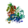
| ||||||||
|---|---|---|---|---|---|---|---|---|---|
| 1 |
| ||||||||
| Unit cell |
| ||||||||
| Details | The biological assembly is a crystallographic dimer. |
-Components
| #1: Protein | Mass: 98017.781 Da / Num. of mol.: 1 Source method: isolated from a genetically manipulated source Source: (gene. exp.) Desulfovibrio desulfuricans (bacteria) / Production host: Bacteria (eubacteria) / References: UniProt: Q9REC4, EC: 1.2.-.- | ||||||
|---|---|---|---|---|---|---|---|
| #2: Chemical | Iron–sulfur cluster  Mass: 175.820 Da / Num. of mol.: 2 / Source method: obtained synthetically / Formula: Fe2S2 Mass: 175.820 Da / Num. of mol.: 2 / Source method: obtained synthetically / Formula: Fe2S2#3: Chemical | ChemComp-2MO / |   Mass: 127.939 Da / Num. of mol.: 1 / Source method: obtained synthetically / Formula: MoO2 Mass: 127.939 Da / Num. of mol.: 1 / Source method: obtained synthetically / Formula: MoO2#4: Chemical | ChemComp-MCN / |   Mass: 696.501 Da / Num. of mol.: 1 / Source method: obtained synthetically / Formula: C19H22N8O13P2S2 Mass: 696.501 Da / Num. of mol.: 1 / Source method: obtained synthetically / Formula: C19H22N8O13P2S2#5: Water | ChemComp-HOH / | Water Mass: 18.015 Da / Num. of mol.: 193 / Source method: isolated from a natural source / Formula: H2O Mass: 18.015 Da / Num. of mol.: 193 / Source method: isolated from a natural source / Formula: H2O |
-Experimental details
-Experiment
| Experiment | Method: X-RAY DIFFRACTION / Number of used crystals: 1 |
|---|
- Sample preparation
| Crystal | Density Matthews: 3.2 Å3/Da / Density % sol: 61.54 % | ||||||||||||||||||||||||||||||
|---|---|---|---|---|---|---|---|---|---|---|---|---|---|---|---|---|---|---|---|---|---|---|---|---|---|---|---|---|---|---|---|
| Crystal grow | Temperature: 298 K / Method: vapor diffusion, hanging drop / pH: 7.6 Details: ammonium sulphate, MES, dioxane, pH 7.6, VAPOR DIFFUSION, HANGING DROP, temperature 298.0K | ||||||||||||||||||||||||||||||
| Crystal grow | *PLUS | ||||||||||||||||||||||||||||||
| Components of the solutions | *PLUS
|
-Data collection
| Diffraction | Mean temperature: 298 K |
|---|---|
| Diffraction source | Source: ROTATING ANODE / Type: ENRAF-NONIUS / Wavelength: 1.542 |
| Detector | Type: MARRESEARCH / Detector: IMAGE PLATE / Date: Jan 1, 1997 |
| Radiation | Protocol: SINGLE WAVELENGTH / Monochromatic (M) / Laue (L): M / Scattering type: x-ray |
| Radiation wavelength | Wavelength: 1.542 Å / Relative weight: 1 |
| Reflection | Resolution: 2.8→24.8 Å / Num. all: 31573 / Num. obs: 190328 / % possible obs: 98.2 % / Observed criterion σ(F): 2 / Observed criterion σ(I): 0 / Redundancy: 10 % / Biso Wilson estimate: 51 Å2 / Rmerge(I) obs: 0.169 / Net I/σ(I): 6.7 |
| Reflection shell | Resolution: 2.8→2.9 Å / Redundancy: 2 % / Rmerge(I) obs: 0.561 / % possible all: 98 |
| Reflection | *PLUS Num. obs: 31573 / Num. measured all: 190328 |
| Reflection shell | *PLUS % possible obs: 98 % / Mean I/σ(I) obs: 2.4 |
- Processing
| Software |
| |||||||||||||||||||||||||
|---|---|---|---|---|---|---|---|---|---|---|---|---|---|---|---|---|---|---|---|---|---|---|---|---|---|---|
| Refinement | Resolution: 2.8→24.8 Å / σ(F): 2 / σ(I): 0 / Stereochemistry target values: Engh & Huber
| |||||||||||||||||||||||||
| Refinement step | Cycle: LAST / Resolution: 2.8→24.8 Å
| |||||||||||||||||||||||||
| Refine LS restraints |
| |||||||||||||||||||||||||
| Software | *PLUS Name: X-PLOR / Version: 3.851 / Classification: refinement | |||||||||||||||||||||||||
| Refinement | *PLUS Highest resolution: 2.8 Å / Lowest resolution: 24.8 Å / Num. reflection obs: 30614 / σ(F): 2 / % reflection Rfree: 3 % / Rfactor obs: 0.164 | |||||||||||||||||||||||||
| Solvent computation | *PLUS | |||||||||||||||||||||||||
| Displacement parameters | *PLUS |
