 Movie
Movie Controller


+ Open data

- Basic information
| Entry | Database: PDB / ID: 1dgg | ||||||
|---|---|---|---|---|---|---|---|
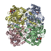
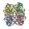
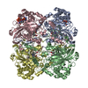
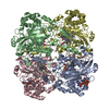
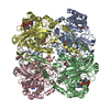
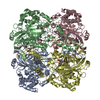
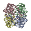
| Title | HUMAN ERYTHROCYTE CATALSE CYANIDE COMPLEX | ||||||
 Components Components | CATALASE | ||||||
 Keywords Keywords | OXIDOREDUCTASE / CATALASE / HEME / NADPH / HYDROGEN PEROXIDE / CYANIDE | ||||||
| Function / homology |  Function and homology information Function and homology informationresponse to phenylpropanoid / aminoacylase activity / catalase complex / hemoglobin metabolic process / response to inactivity / cellular detoxification of hydrogen peroxide / response to light intensity / response to L-ascorbic acid / response to ozone / oxidoreductase activity, acting on peroxide as acceptor ...response to phenylpropanoid / aminoacylase activity / catalase complex / hemoglobin metabolic process / response to inactivity / cellular detoxification of hydrogen peroxide / response to light intensity / response to L-ascorbic acid / response to ozone / oxidoreductase activity, acting on peroxide as acceptor / catalase / UV protection / response to fatty acid / response to vitamin A / catalase activity / triglyceride metabolic process / peroxisomal membrane / ureteric bud development / antioxidant activity / Detoxification of Reactive Oxygen Species / peroxisomal matrix / positive regulation of cell division / response to hyperoxia / response to vitamin E / FOXO-mediated transcription of oxidative stress, metabolic and neuronal genes / response to cadmium ion / aerobic respiration / cholesterol metabolic process / response to reactive oxygen species / response to activity / hydrogen peroxide catabolic process / Peroxisomal protein import / response to lead ion / response to insulin / response to hydrogen peroxide / cellular response to growth factor stimulus / osteoblast differentiation / peroxisome / response to estradiol / NADP binding / response to ethanol / secretory granule lumen / ficolin-1-rich granule lumen / response to hypoxia / positive regulation of phosphatidylinositol 3-kinase/protein kinase B signal transduction / response to xenobiotic stimulus / focal adhesion / intracellular membrane-bounded organelle / heme binding / Neutrophil degranulation / negative regulation of apoptotic process / enzyme binding / protein homodimerization activity / protein-containing complex / mitochondrion / extracellular exosome / extracellular region / membrane / identical protein binding / metal ion binding / cytosol / cytoplasmSimilarity search - Function | ||||||
| Biological species |  Homo sapiens (human) Homo sapiens (human) | ||||||
| Method | X-RAY DIFFRACTION / SYNCHROTRON / Resolution: 1.8 Å | ||||||
 Authors Authors | Putnam, C.D. / Arvai, A.S. / Bourne, Y. / Tainer, J.A. | ||||||
 Citation Citation | Journal: J.Mol.Biol. / Year: 2000 Title: Active and inhibited human catalase structures: ligand and NADPH binding and catalytic mechanism. Authors: Putnam, C.D. / Arvai, A.S. / Bourne, Y. / Tainer, J.A. | ||||||
| History |
|
- Structure visualization
| Structure viewer | Molecule: MolmilJmol/JSmol |
|---|
- Downloads & links
-Download
| PDBx/mmCIF format | 1dgg.cif.gz | 428.8 KB | Display | PDBx/mmCIF format |
|---|---|---|---|---|
| PDB format | pdb1dgg.ent.gz | 348.5 KB | Display | PDB format |
| PDBx/mmJSON format | 1dgg.json.gz | Tree view | PDBx/mmJSON format | |
| Others |  Other downloads Other downloads |
-Validation report
| Arichive directory | https://data.pdbj.org/pub/pdb/validation_reports/dg/1dggftp://data.pdbj.org/pub/pdb/validation_reports/dg/1dgg | HTTPS FTP |
|---|
-Related structure data


-Links
 PDBj
PDBj


- Assembly
| Deposited unit | 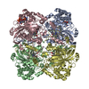
| ||||||||
|---|---|---|---|---|---|---|---|---|---|
| 1 |
| ||||||||
| Unit cell |
|
-Components
| #1: Protein | Mass: 56626.359 Da / Num. of mol.: 4 / Source method: isolated from a natural source / Source: (natural) Homo sapiens (human) / Cell: ERYTHROCYTE / References: UniProt: P04040, catalase#2: Chemical | ChemComp-CYN / Cyanide  Mass: 26.017 Da / Num. of mol.: 4 / Source method: obtained synthetically / Formula: CN Mass: 26.017 Da / Num. of mol.: 4 / Source method: obtained synthetically / Formula: CN#3: Chemical | ChemComp-HEM / Heme B  Mass: 616.487 Da / Num. of mol.: 4 / Source method: obtained synthetically / Formula: C34H32FeN4O4 Mass: 616.487 Da / Num. of mol.: 4 / Source method: obtained synthetically / Formula: C34H32FeN4O4#4: Chemical | Nicotinamide adenine dinucleotide phosphate  Mass: 745.421 Da / Num. of mol.: 2 / Source method: obtained synthetically / Formula: C21H30N7O17P3 Mass: 745.421 Da / Num. of mol.: 2 / Source method: obtained synthetically / Formula: C21H30N7O17P3#5: Water | ChemComp-HOH / | Water Mass: 18.015 Da / Num. of mol.: 1493 / Source method: isolated from a natural source / Formula: H2O Mass: 18.015 Da / Num. of mol.: 1493 / Source method: isolated from a natural source / Formula: H2O |
|---|
-Experimental details
-Experiment
| Experiment | Method: X-RAY DIFFRACTION / Number of used crystals: 1 |
|---|
- Sample preparation
| Crystal | Density Matthews: 3.01 Å3/Da / Density % sol: 59.17 % | ||||||||||||||||||||
|---|---|---|---|---|---|---|---|---|---|---|---|---|---|---|---|---|---|---|---|---|---|
| Crystal grow | Temperature: 298 K / Method: vapor diffusion / pH: 8 Details: 6.5 - 8.0% PEG4000, PROTEIN AT 40 MG/ML IN 50MM TRISCL, PH 8.0, VAPOR DIFFUSION, temperature 298K | ||||||||||||||||||||
| Crystal grow | *PLUS Method: vapor diffusion, hanging drop | ||||||||||||||||||||
| Components of the solutions | *PLUS
|
-Data collection
| Diffraction | Mean temperature: 200 K |
|---|---|
| Diffraction source | Source: SYNCHROTRON / Site: SSRL  / Beamline: BL7-1 / Wavelength: 1.08 / Beamline: BL7-1 / Wavelength: 1.08 |
| Detector | Type: MARRESEARCH / Detector: IMAGE PLATE / Date: Mar 6, 1999 |
| Radiation | Protocol: SINGLE WAVELENGTH / Monochromatic (M) / Laue (L): M / Scattering type: x-ray |
| Radiation wavelength | Wavelength: 1.08 Å / Relative weight: 1 |
| Reflection | Resolution: 1.8→20 Å / Num. all: 196299 / Num. obs: 196299 / % possible obs: 77.7 % / Observed criterion σ(F): 0 / Observed criterion σ(I): -3 / Redundancy: 2.4 % / Biso Wilson estimate: 17.13 Å2 / Rsym value: 0.039 / Net I/σ(I): 18.2 |
| Reflection shell | Resolution: 1.8→1.86 Å / Redundancy: 2.4 % / Rsym value: 0.257 / % possible all: 52.3 |
| Reflection | *PLUS Num. measured all: 473715 / Rmerge(I) obs: 0.039 |
| Reflection shell | *PLUS % possible obs: 52.3 % / Rmerge(I) obs: 0.257 / Mean I/σ(I) obs: 2.6 |
- Processing
| Software |
| ||||||||||||||||||||||||||||
|---|---|---|---|---|---|---|---|---|---|---|---|---|---|---|---|---|---|---|---|---|---|---|---|---|---|---|---|---|---|
| Refinement | Resolution: 1.8→20 Å / σ(F): 0 / σ(I): 0 / Stereochemistry target values: ENGH & HUBER Details: BOND ANGLES AND LENGTHS TO THE HEME IRON FROM THE TYR358 LIGAND, THE CYANIDE LIGAND, AND THE HEME NITROGENS WERE UNCONSTRAINED
| ||||||||||||||||||||||||||||
| Displacement parameters | Biso mean: 42.94 Å2
| ||||||||||||||||||||||||||||
| Refinement step | Cycle: LAST / Resolution: 1.8→20 Å
| ||||||||||||||||||||||||||||
| Refine LS restraints |
| ||||||||||||||||||||||||||||
| Software | *PLUS Name: X-PLOR / Version: 3.851 / Classification: refinement | ||||||||||||||||||||||||||||
| Refinement | *PLUS | ||||||||||||||||||||||||||||
| Solvent computation | *PLUS | ||||||||||||||||||||||||||||
| Displacement parameters | *PLUS | ||||||||||||||||||||||||||||
| Refine LS restraints | *PLUS
|
