 Movie
Movie Controller

[English] 日本語
 Yorodumi
- PDB-1dcf: CRYSTAL STRUCTURE OF THE RECEIVER DOMAIN OF THE ETHYLENE RECEPTOR... -
+ Open data

- Basic information
| Entry | Database: PDB / ID: 1dcf | ||||||
|---|---|---|---|---|---|---|---|
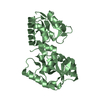
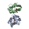
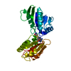
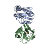
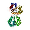
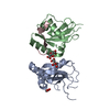
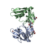
| Title | CRYSTAL STRUCTURE OF THE RECEIVER DOMAIN OF THE ETHYLENE RECEPTOR OF ARABIDOPSIS THALIANA | ||||||
 Components Components | ETR1 PROTEIN | ||||||
 Keywords Keywords |  TRANSFERASE / BETA-ALPHA FIVE SANDWICH TRANSFERASE / BETA-ALPHA FIVE SANDWICH | ||||||
| Function / homology |  Function and homology informationethylene receptor activity / regulation of seedling development / detection of ethylene stimulus / ethylene binding / defense response by callose deposition in cell wall / seed dormancy process / negative regulation of ethylene-activated signaling pathway / sugar mediated signaling pathway / response to gibberellin / response to insect ...ethylene receptor activity / regulation of seedling development / detection of ethylene stimulus / ethylene binding / defense response by callose deposition in cell wall / seed dormancy process / negative regulation of ethylene-activated signaling pathway / sugar mediated signaling pathway / response to gibberellin / response to insect / phloem or xylem histogenesis / response to ethylene / cytokinin metabolic process / regulation of stomatal movement / response to auxin / protein histidine kinase activity / response to abscisic acid / hydrogen peroxide biosynthetic process / histidine kinase / phosphorelay sensor kinase activity / response to salt stress / defense response / response to molecule of bacterial origin / response to heat / defense response to bacterium / cell division / endoplasmic reticulum membrane / endoplasmic reticulum / ATP binding / identical protein binding / metal ion binding Function and homology informationethylene receptor activity / regulation of seedling development / detection of ethylene stimulus / ethylene binding / defense response by callose deposition in cell wall / seed dormancy process / negative regulation of ethylene-activated signaling pathway / sugar mediated signaling pathway / response to gibberellin / response to insect ...ethylene receptor activity / regulation of seedling development / detection of ethylene stimulus / ethylene binding / defense response by callose deposition in cell wall / seed dormancy process / negative regulation of ethylene-activated signaling pathway / sugar mediated signaling pathway / response to gibberellin / response to insect / phloem or xylem histogenesis / response to ethylene / cytokinin metabolic process / regulation of stomatal movement / response to auxin / protein histidine kinase activity / response to abscisic acid / hydrogen peroxide biosynthetic process / histidine kinase / phosphorelay sensor kinase activity / response to salt stress / defense response / response to molecule of bacterial origin / response to heat / defense response to bacterium / cell division / endoplasmic reticulum membrane / endoplasmic reticulum / ATP binding / identical protein binding / metal ion bindingSimilarity search - Function | ||||||
| Biological species |  Arabidopsis thaliana (thale cress) Arabidopsis thaliana (thale cress) | ||||||
| Method | X-RAY DIFFRACTION / Resolution: 2.5 Å | ||||||
 Authors Authors | Muller-Dieckmann, H.J. / Grantz, A. / Kim, S.H. | ||||||
 Citation Citation | Journal: Structure Fold.Des. / Year: 1999 Title: The structure of the signal receiver domain of the Arabidopsis thaliana ethylene receptor ETR1. Authors: Muller-Dieckmann, H.J. / Grantz, A.A. / Kim, S.H. | ||||||
| History |
|
- Structure visualization
| Structure viewer | Molecule: MolmilJmol/JSmol |
|---|
- Downloads & links
-Download
| PDBx/mmCIF format | 1dcf.cif.gz | 37.7 KB | Display | PDBx/mmCIF format |
|---|---|---|---|---|
| PDB format | pdb1dcf.ent.gz | 25.9 KB | Display | PDB format |
| PDBx/mmJSON format | 1dcf.json.gz | Tree view | PDBx/mmJSON format | |
| Others |  Other downloads Other downloads |
-Validation report
| Arichive directory | https://data.pdbj.org/pub/pdb/validation_reports/dc/1dcfftp://data.pdbj.org/pub/pdb/validation_reports/dc/1dcf | HTTPS FTP |
|---|
-Related structure data
| Similar structure data |
|---|


-Links
 PDBj
PDBj


- Assembly
| Deposited unit | 
| ||||||||
|---|---|---|---|---|---|---|---|---|---|
| 1 | 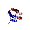
| ||||||||
| Unit cell |
|
-Components
| #1: Protein | Mass: 15304.866 Da / Num. of mol.: 1 / Fragment: RECEIVER DOMAIN Source method: isolated from a genetically manipulated source Source: (gene. exp.) Arabidopsis thaliana (thale cress) / Production host:  Escherichia coli (E. coli) Escherichia coli (E. coli)References: UniProt: P49333, Transferases; Transferring phosphorus-containing groups; Phosphotransferases with a nitrogenous group as acceptor |
|---|---|
| #2: Water | ChemComp-HOH / Water Mass: 18.015 Da / Num. of mol.: 41 / Source method: isolated from a natural source / Formula: H2O Mass: 18.015 Da / Num. of mol.: 41 / Source method: isolated from a natural source / Formula: H2O |
-Experimental details
-Experiment
| Experiment | Method: X-RAY DIFFRACTION / Number of used crystals: 1 |
|---|
- Sample preparation
| Crystal | Density Matthews: 2.1 Å3/Da / Density % sol: 63.4983 % | ||||||||||||||||||||||||||||||
|---|---|---|---|---|---|---|---|---|---|---|---|---|---|---|---|---|---|---|---|---|---|---|---|---|---|---|---|---|---|---|---|
| Crystal grow | Temperature: 277 K / Method: vapor diffusion, hanging drop / pH: 7.4 Details: Li2SO4, HEPES, pH 7.4, VAPOR DIFFUSION, HANGING DROP | ||||||||||||||||||||||||||||||
| Crystal grow | *PLUS pH: 7.5 / Method: vapor diffusion, sitting drop | ||||||||||||||||||||||||||||||
| Components of the solutions | *PLUS
|
-Data collection
| Diffraction | Mean temperature: 120 K |
|---|---|
| Diffraction source | Source: ROTATING ANODE / Type: RIGAKU RU200 / Wavelength: 1.5418 |
| Detector | Type: RIGAKU RAXIS IIC / Detector: IMAGE PLATE / Date: Dec 5, 1996 |
| Radiation | Protocol: SINGLE WAVELENGTH / Monochromatic (M) / Laue (L): M / Scattering type: x-ray |
| Radiation wavelength | Wavelength: 1.5418 Å / Relative weight: 1 |
| Reflection | Resolution: 2.5→100 Å / Num. all: 14608 / Num. obs: 4410 / % possible obs: 87 % / Observed criterion σ(F): 1 / Observed criterion σ(I): 1 / Redundancy: 3.1 % / Biso Wilson estimate: 69 Å2 / Rmerge(I) obs: 0.049 / Net I/σ(I): 13.2 |
| Reflection shell | Resolution: 2.5→2.59 Å / Redundancy: 3 % / Rmerge(I) obs: 0.423 / % possible all: 93.5 |
| Reflection shell | *PLUS % possible obs: 93.5 % |
- Processing
| Software |
| |||||||||||||||||||||||||
|---|---|---|---|---|---|---|---|---|---|---|---|---|---|---|---|---|---|---|---|---|---|---|---|---|---|---|
| Refinement | Resolution: 2.5→500 Å / σ(F): 0 / σ(I): 0 / Stereochemistry target values: engh & huber
| |||||||||||||||||||||||||
| Displacement parameters |
| |||||||||||||||||||||||||
| Refinement step | Cycle: LAST / Resolution: 2.5→500 Å
| |||||||||||||||||||||||||
| Refine LS restraints |
| |||||||||||||||||||||||||
| Xplor file |
| |||||||||||||||||||||||||
| Software | *PLUS Name: 'CNS' / Classification: refinement | |||||||||||||||||||||||||
| Refinement | *PLUS | |||||||||||||||||||||||||
| Solvent computation | *PLUS | |||||||||||||||||||||||||
| Displacement parameters | *PLUS Biso mean: 59 Å2 |
