 Movie
Movie Controller


+ Open data

- Basic information
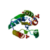
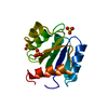
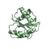
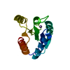
| Entry | Database: PDB / ID: 1d4z | ||||||
|---|---|---|---|---|---|---|---|
| Title | CRYSTAL STRUCTURE OF CHEY-95IV, A HYPERACTIVE CHEY MUTANT | ||||||
 Components Components | CHEMOTAXIS PROTEIN CHEY | ||||||
 Keywords Keywords | SIGNALING PROTEIN / BACTERIAL CHEMOTAXIS / RESPONSE REGULATOR | ||||||
| Function / homology |  Function and homology information Function and homology informationbacterial-type flagellum basal body, C ring / bacterial-type flagellum rotor complex / bacterial-type flagellum-dependent swimming motility / regulation of bacterial-type flagellum-dependent cell motility / aerotaxis / bacterial-type flagellum / regulation of chemotaxis / thermotaxis / internal peptidyl-lysine acetylation / phosphorelay response regulator activity ...bacterial-type flagellum basal body, C ring / bacterial-type flagellum rotor complex / bacterial-type flagellum-dependent swimming motility / regulation of bacterial-type flagellum-dependent cell motility / aerotaxis / bacterial-type flagellum / regulation of chemotaxis / thermotaxis / internal peptidyl-lysine acetylation / phosphorelay response regulator activity / protein acetylation / acetyltransferase activity / phosphorelay signal transduction system / chemotaxis / magnesium ion binding / signal transduction / cytosol / cytoplasmSimilarity search - Function | ||||||
| Biological species |  Escherichia coli (E. coli) Escherichia coli (E. coli) | ||||||
| Method | X-RAY DIFFRACTION / Resolution: 1.9 Å | ||||||
 Authors Authors | Schuster, M. / Zhao, R. / Bourret, R.B. / Collins, E.J. | ||||||
 Citation Citation | Journal: J.Biol.Chem. / Year: 2000 Title: Correlated switch binding and signaling in bacterial chemotaxis. Authors: Schuster, M. / Zhao, R. / Bourret, R.B. / Collins, E.J. | ||||||
| History |
|
- Structure visualization
| Structure viewer | Molecule: MolmilJmol/JSmol |
|---|
- Downloads & links
-Download
| PDBx/mmCIF format | 1d4z.cif.gz | 40.2 KB | Display | PDBx/mmCIF format |
|---|---|---|---|---|
| PDB format | pdb1d4z.ent.gz | 28.1 KB | Display | PDB format |
| PDBx/mmJSON format | 1d4z.json.gz | Tree view | PDBx/mmJSON format | |
| Others |  Other downloads Other downloads |
-Validation report
| Arichive directory | https://data.pdbj.org/pub/pdb/validation_reports/d4/1d4zftp://data.pdbj.org/pub/pdb/validation_reports/d4/1d4z | HTTPS FTP |
|---|
-Related structure data
| Similar structure data |
|---|


-Links
 PDBj
PDBj

- Assembly
| Deposited unit | 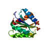
| ||||||||
|---|---|---|---|---|---|---|---|---|---|
| 1 |
| ||||||||
| Unit cell |
|
-Components
| #1: Protein | Mass: 13967.109 Da / Num. of mol.: 1 / Mutation: I95V Source method: isolated from a genetically manipulated source Source: (gene. exp.) Escherichia coli (E. coli) / Production host: Escherichia coli (E. coli) / References: UniProt: P06143, UniProt: P0AE67*PLUS | ||
|---|---|---|---|
| #2: Chemical | Sulfate  Mass: 96.063 Da / Num. of mol.: 3 / Source method: obtained synthetically / Formula: SO4 Mass: 96.063 Da / Num. of mol.: 3 / Source method: obtained synthetically / Formula: SO4#3: Water | ChemComp-HOH / | Water Mass: 18.015 Da / Num. of mol.: 141 / Source method: isolated from a natural source / Formula: H2O Mass: 18.015 Da / Num. of mol.: 141 / Source method: isolated from a natural source / Formula: H2O |
-Experimental details
-Experiment
| Experiment | Method: X-RAY DIFFRACTION / Number of used crystals: 1 |
|---|
- Sample preparation
| Crystal | Density Matthews: 2.02 Å3/Da / Density % sol: 39.19 % | |||||||||||||||||||||||||
|---|---|---|---|---|---|---|---|---|---|---|---|---|---|---|---|---|---|---|---|---|---|---|---|---|---|---|
| Crystal grow | Temperature: 278 K / Method: vapor diffusion, hanging drop / pH: 4.8 Details: AMMONIUM SULPHATE, PEG4000, SODIUM ACETATE, pH 4.8, VAPOR DIFFUSION, HANGING DROP, temperature 278K | |||||||||||||||||||||||||
| Crystal grow | *PLUS Temperature: 4 ℃ / Details: used to seeding | |||||||||||||||||||||||||
| Components of the solutions | *PLUS
|
-Data collection
| Diffraction | Mean temperature: 100 K |
|---|---|
| Diffraction source | Source: ROTATING ANODE / Type: RIGAKU RU200 / Wavelength: 1.5418 |
| Detector | Type: RIGAKU RAXIS IIC / Detector: IMAGE PLATE / Date: Jul 11, 1999 |
| Radiation | Protocol: SINGLE WAVELENGTH / Monochromatic (M) / Laue (L): M / Scattering type: x-ray |
| Radiation wavelength | Wavelength: 1.5418 Å / Relative weight: 1 |
| Reflection | Resolution: 1.9→50 Å / Num. all: 9425 / Num. obs: 9425 / % possible obs: 99.5 % / Observed criterion σ(F): -3 / Observed criterion σ(I): -3 / Redundancy: 11.1 % / Biso Wilson estimate: 13.9 Å2 / Rmerge(I) obs: 0.045 / Net I/σ(I): 44 |
| Reflection shell | Resolution: 1.9→1.93 Å / Redundancy: 6.5 % / Rmerge(I) obs: 0.369 / % possible all: 98 |
| Reflection | *PLUS Num. measured all: 102441 |
| Reflection shell | *PLUS % possible obs: 98 % / Mean I/σ(I) obs: 14 |
- Processing
| Software |
| ||||||||||||||||||||||||||||||||||||||||||||||||||||||||||||
|---|---|---|---|---|---|---|---|---|---|---|---|---|---|---|---|---|---|---|---|---|---|---|---|---|---|---|---|---|---|---|---|---|---|---|---|---|---|---|---|---|---|---|---|---|---|---|---|---|---|---|---|---|---|---|---|---|---|---|---|---|---|
| Refinement | Resolution: 1.9→50 Å / σ(F): 0 / σ(I): 0 / Stereochemistry target values: ENGH AND HUBER
| ||||||||||||||||||||||||||||||||||||||||||||||||||||||||||||
| Refinement step | Cycle: LAST / Resolution: 1.9→50 Å
| ||||||||||||||||||||||||||||||||||||||||||||||||||||||||||||
| Refine LS restraints |
| ||||||||||||||||||||||||||||||||||||||||||||||||||||||||||||
| Software | *PLUS Name: CNS / Classification: refinement | ||||||||||||||||||||||||||||||||||||||||||||||||||||||||||||
| Refinement | *PLUS Highest resolution: 1.9 Å / Lowest resolution: 50 Å / σ(F): 0 / % reflection Rfree: 5 % | ||||||||||||||||||||||||||||||||||||||||||||||||||||||||||||
| Solvent computation | *PLUS | ||||||||||||||||||||||||||||||||||||||||||||||||||||||||||||
| Displacement parameters | *PLUS Biso mean: 12.52 Å2 | ||||||||||||||||||||||||||||||||||||||||||||||||||||||||||||
| Refine LS restraints | *PLUS
| ||||||||||||||||||||||||||||||||||||||||||||||||||||||||||||
| LS refinement shell | *PLUS Highest resolution: 1.9 Å / Lowest resolution: 1.99 Å / Rfactor Rfree: 0.303 / Rfactor obs: 0.257 |
