 Movie
Movie Controller

[English] 日本語
 Yorodumi
- PDB-1d3l: D1D2-ICAM-1 FULLY GLYCOSYLATED, VARIATION OF D1-D2 INTERDOMAIN AN... -
+ Open data

- Basic information
| Entry | Database: PDB / ID: 1d3l | ||||||
|---|---|---|---|---|---|---|---|
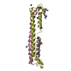
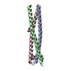
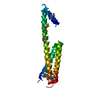
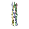
| Title | D1D2-ICAM-1 FULLY GLYCOSYLATED, VARIATION OF D1-D2 INTERDOMAIN ANGLE IN DIFFERENT CRYSTAL STRUCTURES. | ||||||
 Components Components | PROTEIN (INTERCELLULAR ADHESION MOLECULE-1) | ||||||
 Keywords Keywords |  CELL ADHESION / RHINOVIRUS RECEPTOR / ADHESION PROTEIN / GLYCOPROTEIN / IMMUNOGLOBULIN FOLD CELL ADHESION / RHINOVIRUS RECEPTOR / ADHESION PROTEIN / GLYCOPROTEIN / IMMUNOGLOBULIN FOLD | ||||||
| Function / homology |  Function and homology information Function and homology informationregulation of leukocyte mediated cytotoxicity / T cell extravasation / positive regulation of cellular extravasation / regulation of ruffle assembly / T cell antigen processing and presentation / T cell activation via T cell receptor contact with antigen bound to MHC molecule on antigen presenting cell / membrane to membrane docking / adhesion of symbiont to host / establishment of endothelial barrier / cell adhesion mediated by integrin ...regulation of leukocyte mediated cytotoxicity / T cell extravasation / positive regulation of cellular extravasation / regulation of ruffle assembly / T cell antigen processing and presentation / T cell activation via T cell receptor contact with antigen bound to MHC molecule on antigen presenting cell / membrane to membrane docking / adhesion of symbiont to host / establishment of endothelial barrier / cell adhesion mediated by integrin / heterophilic cell-cell adhesion via plasma membrane cell adhesion molecules / leukocyte cell-cell adhesion / leukocyte migration / Interleukin-10 signaling / immunological synapse / Integrin cell surface interactions / negative regulation of endothelial cell apoptotic process / negative regulation of extrinsic apoptotic signaling pathway via death domain receptors / cellular response to leukemia inhibitory factor / cellular response to glucose stimulus / cellular response to amyloid-beta / Immunoregulatory interactions between a Lymphoid and a non-Lymphoid cell / Interferon gamma signaling / transmembrane signaling receptor activity / integrin binding / virus receptor activity / signaling receptor activity / collagen-containing extracellular matrix / Interleukin-4 and Interleukin-13 signaling / receptor-mediated virion attachment to host cell / positive regulation of ERK1 and ERK2 cascade / cell adhesion / membrane raft / external side of plasma membrane / focal adhesion / cell surface / extracellular space / extracellular exosome / membrane / plasma membraneSimilarity search - Function | ||||||
| Biological species |  Homo sapiens (human) Homo sapiens (human) | ||||||
| Method | X-RAY DIFFRACTION / MOLECULAR REPLACEMENT / Resolution: 3.25 Å | ||||||
 Authors Authors | Bella, J. / Kolatkar, P.R. / Rossmann, M.G. | ||||||
 Citation Citation | Journal: EMBO J / Year: 1999 Title: Structural studies of two rhinovirus serotypes complexed with fragments of their cellular receptor. Authors: P R Kolatkar / J Bella / N H Olson / C M Bator / T S Baker / M G Rossmann /  Abstract: Two human rhinovirus serotypes complexed with two- and five-domain soluble fragments of the cellular receptor, intercellular adhesion molecule-1, have been investigated by X-ray crystallographic ...Two human rhinovirus serotypes complexed with two- and five-domain soluble fragments of the cellular receptor, intercellular adhesion molecule-1, have been investigated by X-ray crystallographic analyses of the individual components and by cryo-electron microscopy of the complexes. The three-dimensional image reconstructions provide a molecular envelope within which the crystal structures of the viruses and the receptor fragments can be positioned with accuracy. The N-terminal domain of the receptor binds to the rhinovirus 'canyon' surrounding the icosahedral 5-fold axes. Fitting of molecular models into the image reconstruction density identified the residues on the virus that interact with those on the receptor surface, demonstrating complementarity of the electrostatic patterns for the tip of the N-terminal receptor domain and the floor of the canyon. The complexes seen in the image reconstructions probably represent the first stage of a multistep binding process. A mechanism is proposed for the subsequent viral uncoating process. #1: Journal: Proc.Natl.Acad.Sci.USA / Year: 1998Title: The Structure of the Two Amino-Terminal Domains of Human Icam-1 Suggests How It Functions as a Rhinovirus Receptor and as an Lfa-1 Integrin Ligand. Authors: Bella, J. / Kolatkar, P.R. / Marlor, C.W. / Greve, J.M. / Rossmann, M.G. #2: Journal: Proc.Natl.Acad.Sci.USA / Year: 1998Title: A Dimeric Crystal Structure for the N-Terminal Two Domains of Intercellular Adhesion Molecule-1 Authors: Casasnovas, J.M. / Stehle, T. / Liu, J.H. / Wang, J.H. / Springer, T.A. #3: Journal: J.Mol.Biol. / Year: 1992Title: Preliminary X-Ray Crystallographic Analysis of Intercellular Adhesion Molecule-1 Authors: Kolatkar, P.R. / Oliveira, M.A. / Rossmann, M.G. / Robbins, A.H. / Katti, S. / Hoover-Litty, H. / Forte, C. / Greve, J.M. / Mcclelland, A. / Olson, N.H. | ||||||
| History |
|
- Structure visualization
| Structure viewer | Molecule: MolmilJmol/JSmol |
|---|
- Downloads & links
-Download
| PDBx/mmCIF format | 1d3l.cif.gz | 17.5 KB | Display | PDBx/mmCIF format |
|---|---|---|---|---|
| PDB format | pdb1d3l.ent.gz | 7.9 KB | Display | PDB format |
| PDBx/mmJSON format | 1d3l.json.gz | Tree view | PDBx/mmJSON format | |
| Others |  Other downloads Other downloads |
-Validation report
| Arichive directory | https://data.pdbj.org/pub/pdb/validation_reports/d3/1d3lftp://data.pdbj.org/pub/pdb/validation_reports/d3/1d3l | HTTPS FTP |
|---|
-Related structure data
| Related structure data | 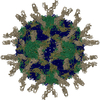 1d3eC  1d3iC 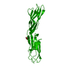 1iamS S: Starting model for refinement C: citing same article ( |
|---|---|
| Similar structure data |


-Links
 PDBj
PDBj


- Assembly
| Deposited unit | 
| ||||||||
|---|---|---|---|---|---|---|---|---|---|
| 1 |
| ||||||||
| Unit cell |
|
-Components
| #1: Protein | Mass: 20438.260 Da / Num. of mol.: 1 / Fragment: FIRST TWO DOMAINS, RESIDUES 1-185 / Source method: isolated from a natural source / Source: (natural) Homo sapiens (human) / References: UniProt: P05362 |
|---|
-Experimental details
-Experiment
| Experiment | Method: X-RAY DIFFRACTION |
|---|
- Sample preparation
| Crystal | Density Matthews: 3.01 Å3/Da / Density % sol: 59.1 % |
|---|---|
| Crystal grow | Details: PROTEIN WAS DESIALATED WITH NEURAMINIDASE (8 HR AT 37 DEGREES IN 100 MM SODIUM ACETATE, PH 6.5, 10 MG/ML PROTEIN, 0.1 ENZYME UNIT/ML), DIALYZED AGAINST 10 MM TRIS, 25 MM NACL (PH 6.0), AND ...Details: PROTEIN WAS DESIALATED WITH NEURAMINIDASE (8 HR AT 37 DEGREES IN 100 MM SODIUM ACETATE, PH 6.5, 10 MG/ML PROTEIN, 0.1 ENZYME UNIT/ML), DIALYZED AGAINST 10 MM TRIS, 25 MM NACL (PH 6.0), AND PASSED THROUGH MONO-Q COLUMN. DESIALATED MATERIAL WAS CRYSTALLIZED BY HANGING DROP METHODS: 17 MG/ML PROTEIN IN BUFFER: 10 MM TRIS,25 MM NACL,1 MM MGCL2,1 MM CACL2, WAS PRECIPITATED FROM 24-27% PEG 3350 IN SAME BUFFER. |
| Crystal grow | *PLUS Method: other / Details: Kolatkar, P.R., (1992) J. Mol. Biol., 225, 1127. |
-Data collection
| Radiation | Protocol: SINGLE WAVELENGTH / Monochromatic (M) / Laue (L): M / Scattering type: x-ray |
|---|---|
| Radiation wavelength | Relative weight: 1 |
| Reflection | Resolution: 2.816→26.582 Å / Num. obs: 4634 / % possible obs: 72.1 % |
| Reflection shell | Resolution: 2.82→2.94 Å / % possible all: 21.8 |
- Processing
| Software | Name: AMoRE / Classification: phasing | ||||||||||||
|---|---|---|---|---|---|---|---|---|---|---|---|---|---|
| Refinement | Method to determine structure: MOLECULAR REPLACEMENT Starting model: PDB ENTRY 1IAM Resolution: 3.25→15 Å / σ(F): 0 / Details: COORDINATES AFTER RIGID-BODY REFINEMENT
| ||||||||||||
| Displacement parameters | Biso mean: 41.89 Å2 | ||||||||||||
| Refinement step | Cycle: LAST / Resolution: 3.25→15 Å
|
