 Movie
Movie Controller


+ Open data

- Basic information
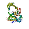
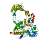
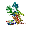
| Entry | Database: PDB / ID: 1cy1 | ||||||
|---|---|---|---|---|---|---|---|
| Title | COMPLEX OF E.COLI DNA TOPOISOMERASE I WITH 5'PTPTPT | ||||||
 Components Components | DNA TOPOISOMERASE I Topoisomerase Topoisomerase | ||||||
 Keywords Keywords | ISOMERASE / DNA TOPOISOMERASE / RELAXING ENZYME | ||||||
| Function / homology |  Function and homology informationDNA topoisomerase activity / DNA topoisomerase / DNA topoisomerase type I (single strand cut, ATP-independent) activity / DNA topological change / chromosome / DNA binding / metal ion binding / cytosol Function and homology informationDNA topoisomerase activity / DNA topoisomerase / DNA topoisomerase type I (single strand cut, ATP-independent) activity / DNA topological change / chromosome / DNA binding / metal ion binding / cytosolSimilarity search - Function | ||||||
| Biological species |  Escherichia coli (E. coli) Escherichia coli (E. coli) | ||||||
| Method | X-RAY DIFFRACTION / SYNCHROTRON / Resolution: 2.3 Å | ||||||
 Authors Authors | Feinberg, H. / Changela, A. / Mondragon, A. | ||||||
 Citation Citation | Journal: Nat.Struct.Biol. / Year: 1999 Title: Protein-nucleotide interactions in E. coli DNA topoisomerase I. Authors: Feinberg, H. / Changela, A. / Mondragon, A. | ||||||
| History |
|
- Structure visualization
| Structure viewer | Molecule: MolmilJmol/JSmol |
|---|
- Downloads & links
-Download
| PDBx/mmCIF format | 1cy1.cif.gz | 121.7 KB | Display | PDBx/mmCIF format |
|---|---|---|---|---|
| PDB format | pdb1cy1.ent.gz | 97.3 KB | Display | PDB format |
| PDBx/mmJSON format | 1cy1.json.gz | Tree view | PDBx/mmJSON format | |
| Others |  Other downloads Other downloads |
-Validation report
| Arichive directory | https://data.pdbj.org/pub/pdb/validation_reports/cy/1cy1ftp://data.pdbj.org/pub/pdb/validation_reports/cy/1cy1 | HTTPS FTP |
|---|
-Related structure data
| Related structure data |  1cy0C  1cy2C  1cy4C  1cy6C  1cy7C  1cy8C C: citing same article ( |
|---|---|
| Similar structure data |


-Links
 PDBj
PDBj


- Assembly
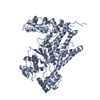
| Deposited unit | 
| ||||||||
|---|---|---|---|---|---|---|---|---|---|
| 1 |
| ||||||||
| Unit cell |
|
-Components
| #1: Protein | Topoisomerase Mass: 67542.500 Da / Num. of mol.: 1 Fragment: 67 KDA N-TERMINAL FRAGMENT OF E.COLI TOPOISOMERASE I Source method: isolated from a genetically manipulated source Source: (gene. exp.) Escherichia coli (E. coli) / Strain: K12 / Plasmid: PGEX-2T / Production host: Escherichia coli (E. coli) / References: UniProt: P06612, DNA topoisomerase | ||||
|---|---|---|---|---|---|
| #2: Chemical | ChemComp-PO4 / Phosphate  Mass: 94.971 Da / Num. of mol.: 1 / Source method: obtained synthetically / Formula: PO4 Mass: 94.971 Da / Num. of mol.: 1 / Source method: obtained synthetically / Formula: PO4 | ||||
| #3: Chemical | 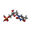  Mass: 322.208 Da / Num. of mol.: 2 / Source method: obtained synthetically / Formula: C10H15N2O8P Mass: 322.208 Da / Num. of mol.: 2 / Source method: obtained synthetically / Formula: C10H15N2O8P#4: Chemical | ChemComp-THP / | Thymidine diphosphate  Type: DNA linking / Mass: 402.188 Da / Num. of mol.: 1 / Source method: obtained synthetically / Formula: C10H16N2O11P2 Type: DNA linking / Mass: 402.188 Da / Num. of mol.: 1 / Source method: obtained synthetically / Formula: C10H16N2O11P2#5: Water | ChemComp-HOH / | Water Mass: 18.015 Da / Num. of mol.: 136 / Source method: isolated from a natural source / Formula: H2O Mass: 18.015 Da / Num. of mol.: 136 / Source method: isolated from a natural source / Formula: H2O |
-Experimental details
-Experiment
| Experiment | Method: X-RAY DIFFRACTION / Number of used crystals: 1 |
|---|
- Sample preparation
| Crystal | Density Matthews: 2.51 Å3/Da / Density % sol: 50.98 % | ||||||||||||||||||||
|---|---|---|---|---|---|---|---|---|---|---|---|---|---|---|---|---|---|---|---|---|---|
| Crystal grow | Temperature: 287 K / Method: vapor diffusion, hanging drop / pH: 5 Details: 2.3M ammonium sulfate, pH 5.0, VAPOR DIFFUSION, HANGING DROP, temperature 287K | ||||||||||||||||||||
| Crystal grow | *PLUS Temperature: 4 ℃ / Details: and 14 degrees centigrade | ||||||||||||||||||||
| Components of the solutions | *PLUS
|
-Data collection
| Diffraction | Mean temperature: 100 K |
|---|---|
| Diffraction source | Source: SYNCHROTRON / Site: CHESS  / Beamline: A1 / Wavelength: 0.9 / Beamline: A1 / Wavelength: 0.9 |
| Detector | Type: PRINCETON 2K / Detector: CCD / Date: Apr 1, 1998 |
| Radiation | Protocol: SINGLE WAVELENGTH / Monochromatic (M) / Laue (L): M / Scattering type: x-ray |
| Radiation wavelength | Wavelength: 0.9 Å / Relative weight: 1 |
| Reflection | Resolution: 2.3→30 Å / Num. all: 423800 / Num. obs: 31029 / % possible obs: 85.8 % / Observed criterion σ(F): 0 / Observed criterion σ(I): 0 / Biso Wilson estimate: 34 Å2 / Rmerge(I) obs: 0.073 |
| Reflection shell | Resolution: 2.3→2.38 Å / Rmerge(I) obs: 0.159 / % possible all: 69.4 |
| Reflection | *PLUS Num. measured all: 423800 |
| Reflection shell | *PLUS % possible obs: 69.4 % |
- Processing
| Software |
| ||||||||||||||||||||||||||||||||||||
|---|---|---|---|---|---|---|---|---|---|---|---|---|---|---|---|---|---|---|---|---|---|---|---|---|---|---|---|---|---|---|---|---|---|---|---|---|---|
| Refinement | Resolution: 2.3→19.98 Å / Rfactor Rfree error: 0.009 / Data cutoff high absF: 1583142.25 / Data cutoff low absF: 0 / Isotropic thermal model: RESTRAINED / Cross valid method: THROUGHOUT / σ(F): 2 Stereochemistry target values: R. A. Engh and R. Huber, Acta Cryst. Sect. A., 1991 Details: Refinement done with REFMAC. The solvent correction was calculated with XPLOR and applied in REFMAC. Final parameters obtained with CNS 0.9
| ||||||||||||||||||||||||||||||||||||
| Solvent computation | Solvent model: FLAT MODEL / Bsol: 18.54 Å2 / ksol: 0.338 e/Å3 | ||||||||||||||||||||||||||||||||||||
| Displacement parameters | Biso mean: 34 Å2
| ||||||||||||||||||||||||||||||||||||
| Refine analyze |
| ||||||||||||||||||||||||||||||||||||
| Refinement step | Cycle: LAST / Resolution: 2.3→19.98 Å
| ||||||||||||||||||||||||||||||||||||
| Refine LS restraints |
| ||||||||||||||||||||||||||||||||||||
| LS refinement shell | Resolution: 2.3→2.44 Å / Rfactor Rfree error: 0.022 / Total num. of bins used: 6
| ||||||||||||||||||||||||||||||||||||
| Xplor file |
| ||||||||||||||||||||||||||||||||||||
| Software | *PLUS Name: 'REFMAC, X-PLOR,CNS 0.9' / Classification: refinement | ||||||||||||||||||||||||||||||||||||
| Refine LS restraints | *PLUS
|
