 Movie
Movie Controller

[English] 日本語
 Yorodumi
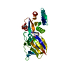
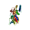
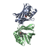
- PDB-1coz: CTP:GLYCEROL-3-PHOSPHATE CYTIDYLYLTRANSFERASE FROM BACILLUS SUBTILIS -
+ Open data

- Basic information
| Entry | Database: PDB / ID: 1coz | ||||||
|---|---|---|---|---|---|---|---|
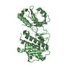
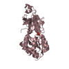
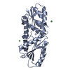
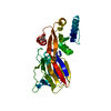
| Title | CTP:GLYCEROL-3-PHOSPHATE CYTIDYLYLTRANSFERASE FROM BACILLUS SUBTILIS | ||||||
 Components Components | PROTEIN (GLYCEROL-3-PHOSPHATE CYTIDYLYLTRANSFERASE) | ||||||
 Keywords Keywords |  TRANSFERASE TRANSFERASE | ||||||
| Function / homology |  Function and homology informationglycerol-3-phosphate cytidylyltransferase / glycerol-3-phosphate cytidylyltransferase activity / teichoic acid biosynthetic process / cell wall organization / metal ion binding / cytoplasm Function and homology informationglycerol-3-phosphate cytidylyltransferase / glycerol-3-phosphate cytidylyltransferase activity / teichoic acid biosynthetic process / cell wall organization / metal ion binding / cytoplasmSimilarity search - Function | ||||||
| Biological species |  Bacillus subtilis (bacteria) Bacillus subtilis (bacteria) | ||||||
| Method | X-RAY DIFFRACTION / MAD / Resolution: 2 Å | ||||||
 Authors Authors | Weber, C.H. / Park, Y.S. / Sanker, S. / Kent, C. / Ludwig, M.L. | ||||||
 Citation Citation | Journal: Structure Fold.Des. / Year: 1999 Title: A prototypical cytidylyltransferase: CTP:glycerol-3-phosphate cytidylyltransferase from bacillus subtilis. Authors: Weber, C.H. / Park, Y.S. / Sanker, S. / Kent, C. / Ludwig, M.L. | ||||||
| History |
|
- Structure visualization
| Structure viewer | Molecule: MolmilJmol/JSmol |
|---|
- Downloads & links
-Download
| PDBx/mmCIF format | 1coz.cif.gz | 68.4 KB | Display | PDBx/mmCIF format |
|---|---|---|---|---|
| PDB format | pdb1coz.ent.gz | 50.9 KB | Display | PDB format |
| PDBx/mmJSON format | 1coz.json.gz | Tree view | PDBx/mmJSON format | |
| Others |  Other downloads Other downloads |
-Validation report
| Arichive directory | https://data.pdbj.org/pub/pdb/validation_reports/co/1cozftp://data.pdbj.org/pub/pdb/validation_reports/co/1coz | HTTPS FTP |
|---|
-Related structure data
| Similar structure data |
|---|


-Links
 PDBj
PDBj
- Assembly
| Deposited unit | 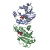
| |||||||||
|---|---|---|---|---|---|---|---|---|---|---|
| 1 |
| |||||||||
| Unit cell |
| |||||||||
| Noncrystallographic symmetry (NCS) | NCS domain:
NCS oper: (Code: given Matrix: (0.9844, 0.1749, 0.0366), Vector : |
-Components
| #1: Protein | Mass: 15295.539 Da / Num. of mol.: 2 Source method: isolated from a genetically manipulated source Source: (gene. exp.) Bacillus subtilis (bacteria) / Strain: BR151-ESC168 / Plasmid: PET-11A / Production host: Escherichia coli (E. coli)References: UniProt: P27623, glycerol-3-phosphate cytidylyltransferase#2: Chemical | Cytidine triphosphate  Mass: 483.156 Da / Num. of mol.: 2 / Source method: obtained synthetically / Formula: C9H16N3O14P3 Mass: 483.156 Da / Num. of mol.: 2 / Source method: obtained synthetically / Formula: C9H16N3O14P3#3: Water | ChemComp-HOH / | Water Mass: 18.015 Da / Num. of mol.: 106 / Source method: isolated from a natural source / Formula: H2O Mass: 18.015 Da / Num. of mol.: 106 / Source method: isolated from a natural source / Formula: H2O |
|---|
-Experimental details
-Experiment
| Experiment | Method: X-RAY DIFFRACTION / Number of used crystals: 1 |
|---|
- Sample preparation
| Crystal | Density Matthews: 2.3 Å3/Da / Density % sol: 48 % | ||||||||||||||||||||||||||||||||||||||||||||||||||
|---|---|---|---|---|---|---|---|---|---|---|---|---|---|---|---|---|---|---|---|---|---|---|---|---|---|---|---|---|---|---|---|---|---|---|---|---|---|---|---|---|---|---|---|---|---|---|---|---|---|---|---|
| Crystal grow | pH: 8.5 Details: 100 MM TRIS PH 8.5 200 MM LITHIUM SULFATE 30% PEG 3350 (BAKER) | ||||||||||||||||||||||||||||||||||||||||||||||||||
| Crystal | *PLUS | ||||||||||||||||||||||||||||||||||||||||||||||||||
| Crystal grow | *PLUS Temperature: 4 ℃ / Method: vapor diffusion, hanging drop | ||||||||||||||||||||||||||||||||||||||||||||||||||
| Components of the solutions | *PLUS
|
-Data collection
| Diffraction | Mean temperature: 277 K |
|---|---|
| Diffraction source | Source: ROTATING ANODE / Type: RIGAKU RU200 / Wavelength: 1.5418 |
| Detector | Type: SDMS / Detector: AREA DETECTOR |
| Radiation | Protocol: SINGLE WAVELENGTH / Monochromatic (M) / Laue (L): M / Scattering type: x-ray |
| Radiation wavelength | Wavelength: 1.5418 Å / Relative weight: 1 |
| Reflection | Resolution: 2→100 Å / Num. obs: 16917 / % possible obs: 88.6 % / Redundancy: 2.68 % / Biso Wilson estimate: 24.7 Å2 / Rsym value: 0.061 |
| Reflection shell | Resolution: 2→2.12 Å / Rsym value: 0.125 / % possible all: 66.4 |
| Reflection | *PLUS Num. measured all: 45342 / Rmerge(I) obs: 0.061 |
| Reflection shell | *PLUS % possible obs: 83.8 % / Rmerge(I) obs: 0.125 |
- Processing
| Software |
| ||||||||||||||||||||||||||||||||||||||||||||||||||||||||||||||||||||||||||||||||
|---|---|---|---|---|---|---|---|---|---|---|---|---|---|---|---|---|---|---|---|---|---|---|---|---|---|---|---|---|---|---|---|---|---|---|---|---|---|---|---|---|---|---|---|---|---|---|---|---|---|---|---|---|---|---|---|---|---|---|---|---|---|---|---|---|---|---|---|---|---|---|---|---|---|---|---|---|---|---|---|---|---|
| Refinement | Method to determine structure: MAD / Resolution: 2→8 Å / Rfactor Rfree error: 0.006 / Data cutoff high absF: 100000 / Data cutoff low absF: 0.001 / Isotropic thermal model: RESTRAINED / Cross valid method: A POSTERIORI / σ(F): 0 Details: BULK SOLVENT MODEL USED GROUP 1 FOR NCS RESTRAINTS: 1 - 37 AND 501 - 537, 47 - 73 AND 547 - 573, 76 - 81 AND 576 - 581, 88 - 93 AND 588 - 593, AND 108 - 126 AND 608 - 626. THE REMAINDER OF ...Details: BULK SOLVENT MODEL USED GROUP 1 FOR NCS RESTRAINTS: 1 - 37 AND 501 - 537, 47 - 73 AND 547 - 573, 76 - 81 AND 576 - 581, 88 - 93 AND 588 - 593, AND 108 - 126 AND 608 - 626. THE REMAINDER OF THE PROTEIN ATOMS WERE IN GROUP 2. THIS P 21 CRYSTAL DISPLAYS MEROHEDRAL TWINNING AND CAN BE INDEXED AS ORTHORHOMBIC C 2 2 21
| ||||||||||||||||||||||||||||||||||||||||||||||||||||||||||||||||||||||||||||||||
| Displacement parameters | Biso mean: 25.2 Å2
| ||||||||||||||||||||||||||||||||||||||||||||||||||||||||||||||||||||||||||||||||
| Refine analyze |
| ||||||||||||||||||||||||||||||||||||||||||||||||||||||||||||||||||||||||||||||||
| Refinement step | Cycle: LAST / Resolution: 2→8 Å
| ||||||||||||||||||||||||||||||||||||||||||||||||||||||||||||||||||||||||||||||||
| Refine LS restraints |
| ||||||||||||||||||||||||||||||||||||||||||||||||||||||||||||||||||||||||||||||||
| Refine LS restraints NCS |
| ||||||||||||||||||||||||||||||||||||||||||||||||||||||||||||||||||||||||||||||||
| LS refinement shell | Resolution: 2→2.12 Å / Rfactor Rfree error: 0.021 / Total num. of bins used: 6
| ||||||||||||||||||||||||||||||||||||||||||||||||||||||||||||||||||||||||||||||||
| Xplor file |
| ||||||||||||||||||||||||||||||||||||||||||||||||||||||||||||||||||||||||||||||||
| Software | *PLUS Name: X-PLOR / Version: 3.851 / Classification: refinement | ||||||||||||||||||||||||||||||||||||||||||||||||||||||||||||||||||||||||||||||||
| Refinement | *PLUS σ(F): 0 / % reflection Rfree: 10 % | ||||||||||||||||||||||||||||||||||||||||||||||||||||||||||||||||||||||||||||||||
| Solvent computation | *PLUS | ||||||||||||||||||||||||||||||||||||||||||||||||||||||||||||||||||||||||||||||||
| Displacement parameters | *PLUS Biso mean: 25.2 Å2 | ||||||||||||||||||||||||||||||||||||||||||||||||||||||||||||||||||||||||||||||||
| Refine LS restraints | *PLUS
| ||||||||||||||||||||||||||||||||||||||||||||||||||||||||||||||||||||||||||||||||
| LS refinement shell | *PLUS Rfactor Rfree: 0.302 / % reflection Rfree: 10.5 % / Rfactor Rwork: 0.271 |
