 Movie
Movie Controller


+ Open data

- Basic information
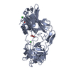
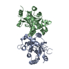
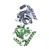
| Entry | Database: PDB / ID: 1cmv | ||||||
|---|---|---|---|---|---|---|---|
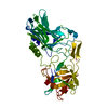
| Title | HUMAN CYTOMEGALOVIRUS PROTEASE | ||||||
 Components Components | HUMAN CYTOMEGALOVIRUS PROTEASE | ||||||
 Keywords Keywords |  SERINE PROTEASE / COAT PROTEIN / HYDROLASE / PHOSPHORYLATION SERINE PROTEASE / COAT PROTEIN / HYDROLASE / PHOSPHORYLATION | ||||||
| Function / homology |  Function and homology informationassemblin / viral release from host cell / host cell cytoplasm / serine-type endopeptidase activity / host cell nucleus / proteolysis / identical protein binding Function and homology informationassemblin / viral release from host cell / host cell cytoplasm / serine-type endopeptidase activity / host cell nucleus / proteolysis / identical protein bindingSimilarity search - Function | ||||||
| Biological species |   Human herpesvirus 5 Human herpesvirus 5 | ||||||
| Method | X-RAY DIFFRACTION / MIR / Resolution: 2.27 Å | ||||||
 Authors Authors | Shieh, H.-S. / Kurumbail, R.G. / Stevens, A.M. / Stegeman, R.A. / Sturman, E.J. / Pak, J.Y. / Wittwer, A.J. / Palmier, M.O. / Wiegand, R.C. / Holwerda, B.C. / Stallings, W.C. | ||||||
 Citation Citation | Journal: Nature / Year: 1996 Title: Three-dimensional structure of human cytomegalovirus protease. Authors: Shieh, H.S. / Kurumbail, R.G. / Stevens, A.M. / Stegeman, R.A. / Sturman, E.J. / Pak, J.Y. / Wittwer, A.J. / Palmier, M.O. / Wiegand, R.C. / Holwerda, B.C. / Stallings, W.C. #1: Journal: J.Biol.Chem. / Year: 1994Title: Activity of Two-Chain Recombinant Human Cytomegalovirus Protease Authors: Holwerda, B.C. / Wittwer, A.J. / Duffin, K.L. / Smith, C. / Toth, M.V. / Carr, L.S. / Wiegand, R.C. / Bryant, M.L. | ||||||
| History |
|
- Structure visualization
| Structure viewer | Molecule: MolmilJmol/JSmol |
|---|
- Downloads & links
-Download
| PDBx/mmCIF format | 1cmv.cif.gz | 106.1 KB | Display | PDBx/mmCIF format |
|---|---|---|---|---|
| PDB format | pdb1cmv.ent.gz | 83.1 KB | Display | PDB format |
| PDBx/mmJSON format | 1cmv.json.gz | Tree view | PDBx/mmJSON format | |
| Others |  Other downloads Other downloads |
-Validation report
| Arichive directory | https://data.pdbj.org/pub/pdb/validation_reports/cm/1cmvftp://data.pdbj.org/pub/pdb/validation_reports/cm/1cmv | HTTPS FTP |
|---|
-Related structure data
| Similar structure data |
|---|


-Links
 PDBj
PDBj- Assembly
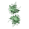
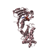
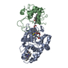
| Deposited unit | 
| ||||||||||||||||||||||||
|---|---|---|---|---|---|---|---|---|---|---|---|---|---|---|---|---|---|---|---|---|---|---|---|---|---|
| 1 |
| ||||||||||||||||||||||||
| Unit cell |
| ||||||||||||||||||||||||
| Noncrystallographic symmetry (NCS) | NCS oper:
|
-Components
| #1: Protein | Mass: 28071.480 Da / Num. of mol.: 2 / Mutation: V141A, A144P Source method: isolated from a genetically manipulated source Source: (gene. exp.) Human herpesvirus 5 / Genus: Cytomegalovirus / Plasmid: PMON9059 / Production host:  Escherichia coli (E. coli) Escherichia coli (E. coli)References: UniProt: P16753, Hydrolases; Acting on peptide bonds (peptidases); Serine endopeptidases#2: Water | ChemComp-HOH / | Water Mass: 18.015 Da / Num. of mol.: 18 / Source method: isolated from a natural source / Formula: H2O Mass: 18.015 Da / Num. of mol.: 18 / Source method: isolated from a natural source / Formula: H2O |
|---|
-Experimental details
-Experiment
| Experiment | Method: X-RAY DIFFRACTION / Number of used crystals: 1 |
|---|
- Sample preparation
| Crystal | Density Matthews: 2.2 Å3/Da / Density % sol: 44 % | ||||||||||||||||||||||||||||||
|---|---|---|---|---|---|---|---|---|---|---|---|---|---|---|---|---|---|---|---|---|---|---|---|---|---|---|---|---|---|---|---|
| Crystal grow | pH: 7.5 Details: 28-30% MONOMETHYL POLYETHYLENE GLYCOL 550 0.05-0.2 M NACL 10% GLYCEROL 50 MM HEPES AT PH 7.5 | ||||||||||||||||||||||||||||||
| Crystal grow | *PLUS Method: vapor diffusion | ||||||||||||||||||||||||||||||
| Components of the solutions | *PLUS
|
-Data collection
| Diffraction | Mean temperature: 296 K |
|---|---|
| Diffraction source | Wavelength: 1.5418 |
| Detector | Type: MARRESEARCH / Detector: IMAGE PLATE / Date: Nov 24, 1995 |
| Radiation | Monochromatic (M) / Laue (L): M / Scattering type: x-ray |
| Radiation wavelength | Wavelength: 1.5418 Å / Relative weight: 1 |
| Reflection | Resolution: 2.27→40 Å / Num. obs: 23705 / % possible obs: 94.5 % / Observed criterion σ(I): -1.5 / Redundancy: 12.5 % / Rmerge(I) obs: 0.08 / Net I/σ(I): 17.38 |
| Reflection shell | Resolution: 2.26→2.34 Å / Rmerge(I) obs: 0.411 / Mean I/σ(I) obs: 1.81 / % possible all: 75.7 |
| Reflection | *PLUS Num. measured all: 297289 |
| Reflection shell | *PLUS % possible obs: 75.7 % |
- Processing
| Software |
| ||||||||||||||||||||||||||||||||||||||||||||||||||||||||||||
|---|---|---|---|---|---|---|---|---|---|---|---|---|---|---|---|---|---|---|---|---|---|---|---|---|---|---|---|---|---|---|---|---|---|---|---|---|---|---|---|---|---|---|---|---|---|---|---|---|---|---|---|---|---|---|---|---|---|---|---|---|---|
| Refinement | Method to determine structure: MIR / Resolution: 2.27→8 Å / σ(F): 2
| ||||||||||||||||||||||||||||||||||||||||||||||||||||||||||||
| Displacement parameters | Biso mean: 26 Å2 | ||||||||||||||||||||||||||||||||||||||||||||||||||||||||||||
| Refinement step | Cycle: LAST / Resolution: 2.27→8 Å
| ||||||||||||||||||||||||||||||||||||||||||||||||||||||||||||
| Refine LS restraints |
| ||||||||||||||||||||||||||||||||||||||||||||||||||||||||||||
| Software | *PLUS Name: X-PLOR / Version: 3.1 / Classification: refinement | ||||||||||||||||||||||||||||||||||||||||||||||||||||||||||||
| Refinement | *PLUS Rfactor Rfree: 0.311 | ||||||||||||||||||||||||||||||||||||||||||||||||||||||||||||
| Solvent computation | *PLUS | ||||||||||||||||||||||||||||||||||||||||||||||||||||||||||||
| Displacement parameters | *PLUS | ||||||||||||||||||||||||||||||||||||||||||||||||||||||||||||
| Refine LS restraints | *PLUS
|
