 Movie
Movie Controller

[English] 日本語
 Yorodumi
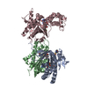
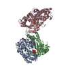
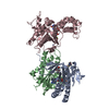
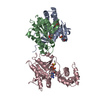
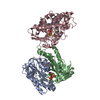
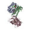
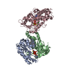
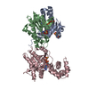
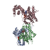
- PDB-1cju: COMPLEX OF GS-ALPHA WITH THE CATALYTIC DOMAINS OF MAMMALIAN ADENY... -
+ Open data

- Basic information
| Entry | Database: PDB / ID: 1cju | ||||||
|---|---|---|---|---|---|---|---|
| Title | COMPLEX OF GS-ALPHA WITH THE CATALYTIC DOMAINS OF MAMMALIAN ADENYLYL CYCLASE: COMPLEX WITH BETA-L-2',3'-DIDEOXYATP AND MG | ||||||
 Components Components |
| ||||||
 Keywords Keywords | LYASE/LYASE/SIGNALING PROTEIN / COMPLEX (LYASE-HYDROLASE) /  HYDROLASE / SIGNAL TRANSDUCING PROTEIN / CYCLASE / EFFECTOR ENZYME / LYASE-LYASE-SIGNALING PROTEIN COMPLEX HYDROLASE / SIGNAL TRANSDUCING PROTEIN / CYCLASE / EFFECTOR ENZYME / LYASE-LYASE-SIGNALING PROTEIN COMPLEX | ||||||
| Function / homology |  Function and homology information Function and homology informationAdenylate cyclase activating pathway / Hedgehog 'off' state / PKA activation / Adenylate cyclase inhibitory pathway / sensory perception of chemical stimulus / adenylate cyclase / mu-type opioid receptor binding / regulation of insulin secretion involved in cellular response to glucose stimulus / corticotropin-releasing hormone receptor 1 binding / cAMP biosynthetic process ...Adenylate cyclase activating pathway / Hedgehog 'off' state / PKA activation / Adenylate cyclase inhibitory pathway / sensory perception of chemical stimulus / adenylate cyclase / mu-type opioid receptor binding / regulation of insulin secretion involved in cellular response to glucose stimulus / corticotropin-releasing hormone receptor 1 binding / cAMP biosynthetic process / G alpha (z) signalling events / adenylate cyclase activity / beta-2 adrenergic receptor binding / cAMP-mediated signaling / adenylate cyclase binding / D1 dopamine receptor binding / positive regulation of cAMP-mediated signaling / adenylate cyclase-activating adrenergic receptor signaling pathway / cellular response to forskolin / ionotropic glutamate receptor binding / adenylate cyclase-inhibiting G protein-coupled receptor signaling pathway / insulin-like growth factor receptor binding / adenylate cyclase activator activity / G-protein beta/gamma-subunit complex binding / adenylate cyclase-modulating G protein-coupled receptor signaling pathway / cilium / adenylate cyclase-activating G protein-coupled receptor signaling pathway / positive regulation of GTPase activity / adenylate cyclase-activating dopamine receptor signaling pathway / heterotrimeric G-protein complex / manganese ion binding / positive regulation of cytosolic calcium ion concentration / intracellular signal transduction / membrane raft / GTPase activity / dendrite / GTP binding / magnesium ion binding / protein-containing complex / ATP binding / membrane / metal ion binding / plasma membrane / cytoplasmSimilarity search - Function | ||||||
| Biological species |  Canis lupus (gray wolf)Rattus norvegicus (Norway rat)Bos taurus (cattle) Canis lupus (gray wolf)Rattus norvegicus (Norway rat)Bos taurus (cattle) | ||||||
| Method | X-RAY DIFFRACTION / SYNCHROTRON / MOLECULAR REPLACEMENT / Resolution: 2.8 Å | ||||||
 Authors Authors | Tesmer, J.J.G. / Sprang, S.R. | ||||||
 Citation Citation | Journal: Science / Year: 1999 Title: Two-metal-Ion catalysis in adenylyl cyclase. Authors: Tesmer, J.J. / Sunahara, R.K. / Johnson, R.A. / Gosselin, G. / Gilman, A.G. / Sprang, S.R. #1: Journal: Science / Year: 1997Title: Crystal Structure of the Catalytic Domains of Adenylyl Cyclase in a Complex with Gsalpha(Dot)Gtpgammas Authors: Tesmer, J.J. / Sunahara, R.K. / Gilman, A.G. / Sprang, S.R. | ||||||
| History |
|
- Structure visualization
| Structure viewer | Molecule: MolmilJmol/JSmol |
|---|
- Downloads & links
-Download
| PDBx/mmCIF format | 1cju.cif.gz | 157.4 KB | Display | PDBx/mmCIF format |
|---|---|---|---|---|
| PDB format | pdb1cju.ent.gz | 123.8 KB | Display | PDB format |
| PDBx/mmJSON format | 1cju.json.gz | Tree view | PDBx/mmJSON format | |
| Others |  Other downloads Other downloads |
-Validation report
| Arichive directory | https://data.pdbj.org/pub/pdb/validation_reports/cj/1cjuftp://data.pdbj.org/pub/pdb/validation_reports/cj/1cju | HTTPS FTP |
|---|
-Related structure data
| Related structure data |  1cjkC  1cjtC  1cjvC  1azsS S: Starting model for refinement C: citing same article ( |
|---|---|
| Similar structure data |

-Links
 PDBj
PDBj


- Assembly
| Deposited unit | 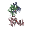
| ||||||||||
|---|---|---|---|---|---|---|---|---|---|---|---|
| 1 |
| ||||||||||
| Unit cell |
|
-Components
-ADENYLATE CYCLASE, TYPE ... , 2 types, 2 molecules AB
| #1: Protein | Adenylyl cyclase / E.C.4.6.1.1 / PROTEIN VC1 Mass: 24495.361 Da / Num. of mol.: 1 / Fragment: C1A DOMAIN OF ADENYLYL CYCLASE / Mutation: V476M Source method: isolated from a genetically manipulated source Source: (gene. exp.) Canis lupus (gray wolf) / Strain: familiaris / Cellular location: CYTOPLASM / Gene: ADENYLYL CYCLASE TYPE V / Organ: CARDIAC MUSCLE / Plasmid: PQE60-H6-VC1(580) / Species (production host): Escherichia coli / Production host:  Escherichia coli BL21(DE3) (bacteria) / Strain (production host): BL21 / Variant (production host): DE3 / References: UniProt: P30803, adenylate cyclase Escherichia coli BL21(DE3) (bacteria) / Strain (production host): BL21 / Variant (production host): DE3 / References: UniProt: P30803, adenylate cyclase |
|---|---|
| #2: Protein | Adenylyl cyclase / E.C.4.6.1.1 / PROTEIN IIC2 / ATP PYROPHOSPHATE-LYASE / ADENYLYL CYCLASE Mass: 23717.033 Da / Num. of mol.: 1 / Fragment: C2A DOMAIN OF ADENYLYL CYCLASE Source method: isolated from a genetically manipulated source Source: (gene. exp.) Rattus norvegicus (Norway rat) / Cellular location: CYTOPLASM / Gene: ADENYLYL CYCLASE TYPE II / Organ: BRAIN / Plasmid: PQE60-ARGC-IIC2 / Species (production host): Escherichia coli / Production host: Escherichia coli BL21(DE3) (bacteria) / Strain (production host): BL21 / Variant (production host): DE3 / References: UniProt: P26769, adenylate cyclase |
-Protein , 1 types, 1 molecules C
| #3: Protein | Mass: 46656.438 Da / Num. of mol.: 1 / Fragment: TRYPSINIZED FRAGMENT / Mutation: N-TERMINAL HEXAHISTIDINE TAG Source method: isolated from a genetically manipulated source Source: (gene. exp.) Bos taurus (cattle) / Cellular location: CYTOPLASM / Gene: GNAS / Plasmid: PQE60-GSALPHA-H / Species (production host): Escherichia coli / Gene (production host): GNAS / Production host: Escherichia coli BL21(DE3) (bacteria) / Strain (production host): BL21 / Variant (production host): DE3 / References: UniProt: P04896 |
|---|
-Non-polymers , 6 types, 49 molecules 

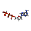


| #4: Chemical |  Mass: 24.305 Da / Num. of mol.: 3 / Source method: obtained synthetically / Formula: Mg Mass: 24.305 Da / Num. of mol.: 3 / Source method: obtained synthetically / Formula: Mg#5: Chemical | ChemComp-FOK / | Forskolin Mass: 410.501 Da / Num. of mol.: 1 / Source method: obtained synthetically / Formula: C22H34O7 Mass: 410.501 Da / Num. of mol.: 1 / Source method: obtained synthetically / Formula: C22H34O7#6: Chemical | ChemComp-DAD / |  Mass: 475.182 Da / Num. of mol.: 1 / Source method: obtained synthetically / Formula: C10H16N5O11P3 Mass: 475.182 Da / Num. of mol.: 1 / Source method: obtained synthetically / Formula: C10H16N5O11P3#7: Chemical | ChemComp-CL / | Chloride Mass: 35.453 Da / Num. of mol.: 1 / Source method: obtained synthetically / Formula: Cl Mass: 35.453 Da / Num. of mol.: 1 / Source method: obtained synthetically / Formula: Cl#8: Chemical | ChemComp-GSP / |  Mass: 539.246 Da / Num. of mol.: 1 / Source method: obtained synthetically / Formula: C10H16N5O13P3S Mass: 539.246 Da / Num. of mol.: 1 / Source method: obtained synthetically / Formula: C10H16N5O13P3S#9: Water | ChemComp-HOH / | WaterMass: 18.015 Da / Num. of mol.: 42 / Source method: isolated from a natural source / Formula: H2O |
|---|
-Experimental details
-Experiment
| Experiment | Method: X-RAY DIFFRACTION / Number of used crystals: 1 |
|---|
- Sample preparation
| Crystal | Density Matthews: 3 Å3/Da / Density % sol: 58 % | ||||||||||||||||||||||||||||||||||||||||||||||||||||||||
|---|---|---|---|---|---|---|---|---|---|---|---|---|---|---|---|---|---|---|---|---|---|---|---|---|---|---|---|---|---|---|---|---|---|---|---|---|---|---|---|---|---|---|---|---|---|---|---|---|---|---|---|---|---|---|---|---|---|
| Crystal grow | Method: vapor diffusion, hanging drop / pH: 5.6 Details: CRYSTALLIZED IN HANGING DROPS CONTAINING PROTEIN MIXED 1:1 WITH WELL SOLUTION OF 7.2-7.5% PEG 8000, 500MM NACL AND 100 MM PHOSPHATE BUFFER (PH 5.4-5.6), VAPOR DIFFUSION, HANGING DROP | ||||||||||||||||||||||||||||||||||||||||||||||||||||||||
| Crystal | *PLUS | ||||||||||||||||||||||||||||||||||||||||||||||||||||||||
| Crystal grow | *PLUS Method: vapor diffusion / Details: Tesmer, J.J., (1997) Science, 278, 1907. / PH range low: 5.6 / PH range high: 5.4 | ||||||||||||||||||||||||||||||||||||||||||||||||||||||||
| Components of the solutions | *PLUS
|
-Data collection
| Diffraction | Mean temperature: 100 K |
|---|---|
| Diffraction source | Source: SYNCHROTRON / Site: CHESS  / Beamline: F2 / Wavelength: 0.923 / Beamline: F2 / Wavelength: 0.923 |
| Detector | Type: ADSC QUANTUM 4 / Detector: CCD / Date: Aug 15, 1998 |
| Radiation | Protocol: SINGLE WAVELENGTH / Monochromatic (M) / Laue (L): M / Scattering type: x-ray |
| Radiation wavelength | Wavelength: 0.923 Å / Relative weight: 1 |
| Reflection | Resolution: 2.8→40 Å / Num. obs: 22654 / % possible obs: 80.2 % / Observed criterion σ(I): -3 / Redundancy: 3.8 % / Biso Wilson estimate: 40.3 Å2 / Rsym value: 0.136 / Net I/σ(I): 8.3 |
| Reflection shell | Resolution: 2.8→2.95 Å / Redundancy: 2.6 % / Mean I/σ(I) obs: 2.8 / Rsym value: 0.299 / % possible all: 72.1 |
| Reflection | *PLUS Rmerge(I) obs: 0.136 |
- Processing
| Software |
| ||||||||||||||||||||||||||||||||||||||||||||||||||||||||||||||||||||||||||||||||
|---|---|---|---|---|---|---|---|---|---|---|---|---|---|---|---|---|---|---|---|---|---|---|---|---|---|---|---|---|---|---|---|---|---|---|---|---|---|---|---|---|---|---|---|---|---|---|---|---|---|---|---|---|---|---|---|---|---|---|---|---|---|---|---|---|---|---|---|---|---|---|---|---|---|---|---|---|---|---|---|---|---|
| Refinement | Method to determine structure: MOLECULAR REPLACEMENT Starting model: PDB ENTRY 1AZS Resolution: 2.8→15 Å / Rfactor Rfree error: 0.006 / Data cutoff high rms absF: 1544755.45 / Isotropic thermal model: RESTRAINED / Cross valid method: THROUGHOUT / σ(F): 0 Details: A BULK SOLVENT CORRECTION WAS USED REFLECTIONS WITH L>21 WERE OMITTED FROM REFINEMENT
| ||||||||||||||||||||||||||||||||||||||||||||||||||||||||||||||||||||||||||||||||
| Solvent computation | Solvent model: FLAT MODEL / Bsol: 10 Å2 / ksol: 0.2822 e/Å3 | ||||||||||||||||||||||||||||||||||||||||||||||||||||||||||||||||||||||||||||||||
| Displacement parameters | Biso mean: 43 Å2
| ||||||||||||||||||||||||||||||||||||||||||||||||||||||||||||||||||||||||||||||||
| Refine analyze |
| ||||||||||||||||||||||||||||||||||||||||||||||||||||||||||||||||||||||||||||||||
| Refinement step | Cycle: LAST / Resolution: 2.8→15 Å
| ||||||||||||||||||||||||||||||||||||||||||||||||||||||||||||||||||||||||||||||||
| Refine LS restraints |
| ||||||||||||||||||||||||||||||||||||||||||||||||||||||||||||||||||||||||||||||||
| LS refinement shell | Resolution: 2.8→2.9 Å / Rfactor Rfree error: 0.031 / Total num. of bins used: 10
| ||||||||||||||||||||||||||||||||||||||||||||||||||||||||||||||||||||||||||||||||
| Xplor file |
| ||||||||||||||||||||||||||||||||||||||||||||||||||||||||||||||||||||||||||||||||
| Software | *PLUS Name: CNS / Version: 0.5 / Classification: refinement | ||||||||||||||||||||||||||||||||||||||||||||||||||||||||||||||||||||||||||||||||
| Refinement | *PLUS σ(F): 0 / % reflection Rfree: 10 % / Rfactor obs: 0.222 | ||||||||||||||||||||||||||||||||||||||||||||||||||||||||||||||||||||||||||||||||
| Solvent computation | *PLUS | ||||||||||||||||||||||||||||||||||||||||||||||||||||||||||||||||||||||||||||||||
| Displacement parameters | *PLUS Biso mean: 43 Å2 | ||||||||||||||||||||||||||||||||||||||||||||||||||||||||||||||||||||||||||||||||
| Refine LS restraints | *PLUS
| ||||||||||||||||||||||||||||||||||||||||||||||||||||||||||||||||||||||||||||||||
| LS refinement shell | *PLUS Rfactor Rfree: 0.403 / % reflection Rfree: 10.3 % / Rfactor Rwork: 0.352 |
