 Movie
Movie Controller

[English] 日本語
 Yorodumi
- PDB-1cjd: THE BACTERIOPHAGE PRD1 COAT PROTEIN, P3, IS STRUCTURALLY SIMILAR ... -
+ Open data

- Basic information
| Entry | Database: PDB / ID: 1cjd | ||||||
|---|---|---|---|---|---|---|---|
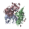
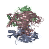
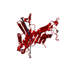
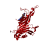
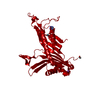
| Title | THE BACTERIOPHAGE PRD1 COAT PROTEIN, P3, IS STRUCTURALLY SIMILAR TO HUMAN ADENOVIRUS HEXON | ||||||
 Components Components | PROTEIN (MAJOR CAPSID PROTEIN (P3)) | ||||||
 Keywords Keywords |  VIRAL PROTEIN / BACTERIOPHAGE PRD1 / COAT PROTEIN / JELLY ROLL VIRAL PROTEIN / BACTERIOPHAGE PRD1 / COAT PROTEIN / JELLY ROLL | ||||||
| Function / homology |  Function and homology information Function and homology information | ||||||
| Biological species |   Enterobacteria phage PRD1 (virus) Enterobacteria phage PRD1 (virus) | ||||||
| Method | X-RAY DIFFRACTION / SYNCHROTRON / MAD / Resolution: 1.85 Å | ||||||
 Authors Authors | Benson, S.D. / Bamford, J.K.H. / Bamford, D.H. / Burnett, R.M. | ||||||
 Citation Citation | Journal: Cell / Year: 1999 Title: Viral evolution revealed by bacteriophage PRD1 and human adenovirus coat protein structures. Authors: S D Benson / J K Bamford / D H Bamford / R M Burnett /  Abstract: The unusual bacteriophage PRD1 features a membrane beneath its icosahedral protein coat. The crystal structure of the major coat protein, P3, at 1.85 A resolution reveals a molecule with three ...The unusual bacteriophage PRD1 features a membrane beneath its icosahedral protein coat. The crystal structure of the major coat protein, P3, at 1.85 A resolution reveals a molecule with three interlocking subunits, each with two eight-stranded viral jelly rolls normal to the viral capsid, and putative membrane-interacting regions. Surprisingly, the P3 molecule closely resembles hexon, the equivalent protein in human adenovirus. Both viruses also have similar overall architecture, with identical capsid lattices and attachment proteins at their vertices. Although these two dsDNA viruses infect hosts from very different kingdoms, their striking similarities, from major coat protein through capsid architecture, strongly suggest their evolutionary relationship. #1: Journal: J.Mol.Biol. / Year: 1993 Title: Crystallization of the major coat protein of PRD1, a bacteriophage with an internal membrane. Authors: Stewart, P.L. / Ghosh, S. / Bamford, D.H. / Burnett, R.M. #2: Journal: Virology / Year: 1990 Title: Capsomer proteins of bacteriophage PRD1, a bacterial virus with a membrane. Authors: Bamford, J.K. / Bamford, D.H. | ||||||
| History |
|
- Structure visualization
| Structure viewer | Molecule: MolmilJmol/JSmol |
|---|
- Downloads & links
-Download
| PDBx/mmCIF format | 1cjd.cif.gz | 224.6 KB | Display | PDBx/mmCIF format |
|---|---|---|---|---|
| PDB format | pdb1cjd.ent.gz | 182.5 KB | Display | PDB format |
| PDBx/mmJSON format | 1cjd.json.gz | Tree view | PDBx/mmJSON format | |
| Others |  Other downloads Other downloads |
-Validation report
| Arichive directory | https://data.pdbj.org/pub/pdb/validation_reports/cj/1cjdftp://data.pdbj.org/pub/pdb/validation_reports/cj/1cjd | HTTPS FTP |
|---|
-Related structure data
| Similar structure data |
|---|


-Links
 PDBj
PDBj

- Assembly
| Deposited unit | 
| ||||||||||||
|---|---|---|---|---|---|---|---|---|---|---|---|---|---|
| 1 |
| ||||||||||||
| Unit cell |
| ||||||||||||
| Noncrystallographic symmetry (NCS) | NCS oper:
|
-Components
| #1: Protein | Mass: 43346.219 Da / Num. of mol.: 3 Source method: isolated from a genetically manipulated source Source: (gene. exp.) Enterobacteria phage PRD1 (virus) / Genus: Tectivirus / Production host:  Salmonella typhimurium (bacteria) / References: UniProt: P22535 Salmonella typhimurium (bacteria) / References: UniProt: P22535#2: Water | ChemComp-HOH / | Water Mass: 18.015 Da / Num. of mol.: 473 / Source method: isolated from a natural source / Formula: H2O Mass: 18.015 Da / Num. of mol.: 473 / Source method: isolated from a natural source / Formula: H2O |
|---|
-Experimental details
-Experiment
| Experiment | Method: X-RAY DIFFRACTION / Number of used crystals: 2 |
|---|
- Sample preparation
| Crystal | Density Matthews: 3.8 Å3/Da / Density % sol: 64 % Description: MAD PHASES WERE COLLECTED ON A SE-MET P3 DERIVATIVE AND THE MODEL USED TO PHASE THE NATIVE P3 DATA | ||||||||||||||||||||||||||||||||||||||||||
|---|---|---|---|---|---|---|---|---|---|---|---|---|---|---|---|---|---|---|---|---|---|---|---|---|---|---|---|---|---|---|---|---|---|---|---|---|---|---|---|---|---|---|---|
| Crystal grow | pH: 4.2 Details: 35% (W/V) MPD, 0.2 M NACL, 0.1 M SODIUM ACETATE, PH 4.2 | ||||||||||||||||||||||||||||||||||||||||||
| Crystal | *PLUS | ||||||||||||||||||||||||||||||||||||||||||
| Crystal grow | *PLUS pH: 8.5 / Method: vapor diffusion, hanging drop | ||||||||||||||||||||||||||||||||||||||||||
| Components of the solutions | *PLUS
|
-Data collection
| Diffraction | Mean temperature: 100 K | ||||||||||||
|---|---|---|---|---|---|---|---|---|---|---|---|---|---|
| Diffraction source | Source: SYNCHROTRON / Site: NSLS / Beamline: X12C / Wavelength: 0.9500, 0.9786, 0.9790 | ||||||||||||
| Detector | Type: BRANDEIS / Detector: CCD / Date: Jun 1, 1997 / Details: MIRRORS | ||||||||||||
| Radiation | Monochromator: SI(111) / Protocol: MAD / Monochromatic (M) / Laue (L): M / Scattering type: x-ray | ||||||||||||
| Radiation wavelength |
| ||||||||||||
| Reflection | Resolution: 1.85→35 Å / Num. obs: 151224 / % possible obs: 99.8 % / Redundancy: 5.2 % / Biso Wilson estimate: 10.1 Å2 / Rsym value: 0.046 / Net I/σ(I): 12 | ||||||||||||
| Reflection shell | Resolution: 1.8→1.9 Å / Redundancy: 2 % / Mean I/σ(I) obs: 9 / Rsym value: 0.197 / % possible all: 91.6 | ||||||||||||
| Reflection | *PLUS Rmerge(I) obs: 0.046 |
- Processing
| Software |
| ||||||||||||||||||||||||||||||||||||||||||||||||||||||||||||||||||||||||||||||||
|---|---|---|---|---|---|---|---|---|---|---|---|---|---|---|---|---|---|---|---|---|---|---|---|---|---|---|---|---|---|---|---|---|---|---|---|---|---|---|---|---|---|---|---|---|---|---|---|---|---|---|---|---|---|---|---|---|---|---|---|---|---|---|---|---|---|---|---|---|---|---|---|---|---|---|---|---|---|---|---|---|---|
| Refinement | Method to determine structure: MAD / Resolution: 1.85→35 Å / Rfactor Rfree error: 0.006 / Data cutoff high absF: 10000000 / Data cutoff low absF: 0.001 / Isotropic thermal model: RESTRAINED / Cross valid method: THROUGHOUT / σ(F): 2 / Details: BULK SOLVENT MODEL USED
| ||||||||||||||||||||||||||||||||||||||||||||||||||||||||||||||||||||||||||||||||
| Displacement parameters | Biso mean: 25.9 Å2
| ||||||||||||||||||||||||||||||||||||||||||||||||||||||||||||||||||||||||||||||||
| Refine analyze |
| ||||||||||||||||||||||||||||||||||||||||||||||||||||||||||||||||||||||||||||||||
| Refinement step | Cycle: LAST / Resolution: 1.85→35 Å
| ||||||||||||||||||||||||||||||||||||||||||||||||||||||||||||||||||||||||||||||||
| Refine LS restraints |
| ||||||||||||||||||||||||||||||||||||||||||||||||||||||||||||||||||||||||||||||||
| Refine LS restraints NCS | NCS model details: UNRESTRAINED | ||||||||||||||||||||||||||||||||||||||||||||||||||||||||||||||||||||||||||||||||
| LS refinement shell | Resolution: 1.85→1.93 Å / Rfactor Rfree error: 0.02 / Total num. of bins used: 8
| ||||||||||||||||||||||||||||||||||||||||||||||||||||||||||||||||||||||||||||||||
| Xplor file |
| ||||||||||||||||||||||||||||||||||||||||||||||||||||||||||||||||||||||||||||||||
| Software | *PLUS Name: X-PLOR / Version: 3.851 / Classification: refinement | ||||||||||||||||||||||||||||||||||||||||||||||||||||||||||||||||||||||||||||||||
| Refinement | *PLUS Lowest resolution: 35 Å / σ(F): 2 / % reflection Rfree: 1.1 % / Rfactor obs: 0.178 | ||||||||||||||||||||||||||||||||||||||||||||||||||||||||||||||||||||||||||||||||
| Solvent computation | *PLUS | ||||||||||||||||||||||||||||||||||||||||||||||||||||||||||||||||||||||||||||||||
| Displacement parameters | *PLUS Biso mean: 25.9 Å2 | ||||||||||||||||||||||||||||||||||||||||||||||||||||||||||||||||||||||||||||||||
| Refine LS restraints | *PLUS
| ||||||||||||||||||||||||||||||||||||||||||||||||||||||||||||||||||||||||||||||||
| LS refinement shell | *PLUS Rfactor Rfree: 0.2 / % reflection Rfree: 1.1 % / Rfactor Rwork: 0.196 |
