 Movie
Movie Controller

[English] 日本語
 Yorodumi
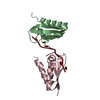
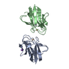
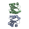
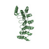
- PDB-1cid: CRYSTAL STRUCTURE OF DOMAINS 3 & 4 OF RAT CD4 AND THEIR RELATIONS... -
+ Open data

- Basic information
| Entry | Database: PDB / ID: 1cid | ||||||
|---|---|---|---|---|---|---|---|
| Title | CRYSTAL STRUCTURE OF DOMAINS 3 & 4 OF RAT CD4 AND THEIR RELATIONSHIP TO THE NH2-TERMINAL DOMAINS | ||||||
 Components Components | T CELL SURFACE GLYCOPROTEIN CD4 | ||||||
 Keywords Keywords | T-CELL SURFACE GLYCOPROTEIN | ||||||
| Function / homology |  Function and homology information Function and homology informationTranslocation of ZAP-70 to Immunological synapse / induction by virus of host cell-cell fusion / PD-1 signaling / Phosphorylation of CD3 and TCR zeta chains / cytokine production / Downstream TCR signaling / Generation of second messenger molecules / Alpha-defensins / Other interleukin signaling / helper T cell enhancement of adaptive immune response ...Translocation of ZAP-70 to Immunological synapse / induction by virus of host cell-cell fusion / PD-1 signaling / Phosphorylation of CD3 and TCR zeta chains / cytokine production / Downstream TCR signaling / Generation of second messenger molecules / Alpha-defensins / Other interleukin signaling / helper T cell enhancement of adaptive immune response /  interleukin-16 binding / interleukin-16 receptor activity / maintenance of protein location in cell / T cell selection / Cargo recognition for clathrin-mediated endocytosis / Clathrin-mediated endocytosis / MHC class II protein binding / response to vitamin D / cellular response to granulocyte macrophage colony-stimulating factor stimulus / interleukin-15-mediated signaling pathway / positive regulation of monocyte differentiation / positive regulation of kinase activity / regulation of T cell activation / positive regulation of calcium ion transport into cytosol / plasma membrane => GO:0005886 / immunoglobulin binding / regulation of calcium ion transport / macrophage differentiation / T cell differentiation / positive regulation of protein kinase activity / coreceptor activity / positive regulation of calcium-mediated signaling / positive regulation of T cell proliferation / protein tyrosine kinase binding / T cell activation / positive regulation of T cell activation / positive regulation of peptidyl-tyrosine phosphorylation / response to estradiol / positive regulation of canonical NF-kappaB signal transduction / defense response to Gram-negative bacterium / positive regulation of MAPK cascade / adaptive immune response / positive regulation of viral entry into host cell / positive regulation of ERK1 and ERK2 cascade / cell surface receptor signaling pathway / cell adhesion / positive regulation of protein phosphorylation / membrane raft / endoplasmic reticulum lumen / external side of plasma membrane / signaling receptor binding / endoplasmic reticulum membrane / protein kinase binding / positive regulation of DNA-templated transcription / enzyme binding / cell surface / signal transduction / protein homodimerization activity / zinc ion binding / identical protein binding / plasma membrane interleukin-16 binding / interleukin-16 receptor activity / maintenance of protein location in cell / T cell selection / Cargo recognition for clathrin-mediated endocytosis / Clathrin-mediated endocytosis / MHC class II protein binding / response to vitamin D / cellular response to granulocyte macrophage colony-stimulating factor stimulus / interleukin-15-mediated signaling pathway / positive regulation of monocyte differentiation / positive regulation of kinase activity / regulation of T cell activation / positive regulation of calcium ion transport into cytosol / plasma membrane => GO:0005886 / immunoglobulin binding / regulation of calcium ion transport / macrophage differentiation / T cell differentiation / positive regulation of protein kinase activity / coreceptor activity / positive regulation of calcium-mediated signaling / positive regulation of T cell proliferation / protein tyrosine kinase binding / T cell activation / positive regulation of T cell activation / positive regulation of peptidyl-tyrosine phosphorylation / response to estradiol / positive regulation of canonical NF-kappaB signal transduction / defense response to Gram-negative bacterium / positive regulation of MAPK cascade / adaptive immune response / positive regulation of viral entry into host cell / positive regulation of ERK1 and ERK2 cascade / cell surface receptor signaling pathway / cell adhesion / positive regulation of protein phosphorylation / membrane raft / endoplasmic reticulum lumen / external side of plasma membrane / signaling receptor binding / endoplasmic reticulum membrane / protein kinase binding / positive regulation of DNA-templated transcription / enzyme binding / cell surface / signal transduction / protein homodimerization activity / zinc ion binding / identical protein binding / plasma membraneSimilarity search - Function | ||||||
| Biological species |  Rattus norvegicus (Norway rat) Rattus norvegicus (Norway rat) | ||||||
| Method | X-RAY DIFFRACTION / Resolution: 2.8 Å | ||||||
 Authors Authors | Brady, R.L. / Dodson, E.J. / Lange, G. | ||||||
 Citation Citation | Journal: Science / Year: 1993 Title: Crystal structure of domains 3 and 4 of rat CD4: relation to the NH2-terminal domains. Authors: Brady, R.L. / Dodson, E.J. / Dodson, G.G. / Lange, G. / Davis, S.J. / Williams, A.F. / Barclay, A.N. | ||||||
| History |
|
- Structure visualization
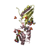
| Structure viewer | Molecule: MolmilJmol/JSmol |
|---|
- Downloads & links
-Download
| PDBx/mmCIF format | 1cid.cif.gz | 44.9 KB | Display | PDBx/mmCIF format |
|---|---|---|---|---|
| PDB format | pdb1cid.ent.gz | 34 KB | Display | PDB format |
| PDBx/mmJSON format | 1cid.json.gz | Tree view | PDBx/mmJSON format | |
| Others |  Other downloads Other downloads |
-Validation report
| Arichive directory | https://data.pdbj.org/pub/pdb/validation_reports/ci/1cidftp://data.pdbj.org/pub/pdb/validation_reports/ci/1cid | HTTPS FTP |
|---|
-Related structure data
| Similar structure data |
|---|


-Links
 PDBj
PDBj


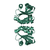
- Assembly
| Deposited unit | 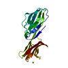
| ||||||||
|---|---|---|---|---|---|---|---|---|---|
| 1 |
| ||||||||
| Unit cell |
| ||||||||
| Atom site foot note | 1: SULFATE OXYGENS ARE NOT VISIBLE IN ELECTRON DENSITY MAP. / 2: RESIDUES PRO 62 AND PRO 71 ARE CIS PROLINES. |
-Components
| #1: Protein | Mass: 19706.373 Da / Num. of mol.: 1 Source method: isolated from a genetically manipulated source Source: (gene. exp.) Rattus norvegicus (Norway rat) / Organ: OVARY / References: UniProt: P05540 |
|---|---|
| #2: Chemical | ChemComp-SO4 / Sulfate  Mass: 96.063 Da / Num. of mol.: 1 / Source method: obtained synthetically / Formula: SO4 Mass: 96.063 Da / Num. of mol.: 1 / Source method: obtained synthetically / Formula: SO4 |
| #3: Water | ChemComp-HOH / Water Mass: 18.015 Da / Num. of mol.: 14 / Source method: isolated from a natural source / Formula: H2O Mass: 18.015 Da / Num. of mol.: 14 / Source method: isolated from a natural source / Formula: H2O |
-Experimental details
-Experiment
| Experiment | Method: X-RAY DIFFRACTION |
|---|
- Sample preparation
| Crystal | Density Matthews: 3.66 Å3/Da / Density % sol: 66.35 % | |||||||||||||||||||||||||
|---|---|---|---|---|---|---|---|---|---|---|---|---|---|---|---|---|---|---|---|---|---|---|---|---|---|---|
| Crystal grow | *PLUS pH: 8 / Method: vapor diffusion, sitting drop | |||||||||||||||||||||||||
| Components of the solutions | *PLUS
|
-Data collection
| Radiation | Scattering type: x-ray |
|---|---|
| Radiation wavelength | Relative weight: 1 |
| Reflection | *PLUS Highest resolution: 2.8 Å / Num. obs: 7274 / % possible obs: 99.1 % / Redundancy: 5.7 % / Num. measured all: 41516 / Rmerge(I) obs: 0.065 |
- Processing
| Software | Name: PROLSQ / Classification: refinement | ||||||||||||
|---|---|---|---|---|---|---|---|---|---|---|---|---|---|
| Refinement | Resolution: 2.8→10 Å / Rfactor all: 0.233 | ||||||||||||
| Refinement step | Cycle: LAST / Resolution: 2.8→10 Å
| ||||||||||||
| Refine LS restraints |
| ||||||||||||
| Refinement | *PLUS Highest resolution: 2.8 Å / Lowest resolution: 10 Å / Num. reflection obs: 7133 / Rfactor obs: 0.233 | ||||||||||||
| Solvent computation | *PLUS | ||||||||||||
| Displacement parameters | *PLUS |
