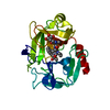
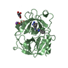
Mass: 25484.211 Da / Num. of mol.: 1 / Source method: isolated from a natural source / Source: (natural) Homo sapiens (human) / References: UniProt: P08311, cathepsin G
Highest resolution: 1.8 Å / Num. measured all: 68129
Reflection shell
*PLUS
Highest resolution: 1.8 Å / Lowest resolution: 1.86 Å / % possible obs: 95.3 % / Rmerge(I) obs: 0.327
-
Processing
Software
Name
Version
Classification
MOSFLM
datareduction
ROTAVATA
datareduction
Agrovata
datareduction
X-PLOR
3.1
modelbuilding
X-PLOR
3.1
refinement
CCP4
(AGROVATA
datascaling
ROTAVATA
datascaling
X-PLOR
3.1
phasing
Refinement
Method to determine structure: MOLECULAR REPLACEMENT Starting model: BOVINE BETA TRYPSIN (W.BODE) Resolution: 1.8→6 Å / σ(F): 0 Details: RESIDUES SER A 36A AND PRO A 36B ARE NOT DEFINED IN ELECTRON DENSITY AND THUS NOT WEIGHTED IN THE REFINEMENT. RESIDUES SER A 36A AND PRO A 36B ARE NOT DEFINED IN ELECTRON DENSITY AND THUS ...Details: RESIDUES SER A 36A AND PRO A 36B ARE NOT DEFINED IN ELECTRON DENSITY AND THUS NOT WEIGHTED IN THE REFINEMENT. RESIDUES SER A 36A AND PRO A 36B ARE NOT DEFINED IN ELECTRON DENSITY AND THUS NOT WEIGHTED IN THE REFINEMENT.
Rfactor
Num. reflection
% reflection
Rfree
0.24
-
10 %
Rwork
0.19
-
-
obs
0.19
18433
94.2 %
Displacement parameters
Biso mean: 22.43 Å2
Refinement step
Cycle: LAST / Resolution: 1.8→6 Å
Protein
Nucleic acid
Ligand
Solvent
Total
Num. atoms
1786
0
33
145
1964
Refine LS restraints
Refine-ID
Type
Dev ideal
X-RAY DIFFRACTION
x_bond_d
0.013
X-RAY DIFFRACTION
x_bond_d_na
X-RAY DIFFRACTION
x_bond_d_prot
X-RAY DIFFRACTION
x_angle_d
X-RAY DIFFRACTION
x_angle_d_na
X-RAY DIFFRACTION
x_angle_d_prot
X-RAY DIFFRACTION
x_angle_deg
1.765
X-RAY DIFFRACTION
x_angle_deg_na
X-RAY DIFFRACTION
x_angle_deg_prot
X-RAY DIFFRACTION
x_dihedral_angle_d
X-RAY DIFFRACTION
x_dihedral_angle_d_na
X-RAY DIFFRACTION
x_dihedral_angle_d_prot
X-RAY DIFFRACTION
x_improper_angle_d
X-RAY DIFFRACTION
x_improper_angle_d_na
X-RAY DIFFRACTION
x_improper_angle_d_prot
X-RAY DIFFRACTION
x_mcbond_it
2.05
X-RAY DIFFRACTION
x_mcangle_it
X-RAY DIFFRACTION
x_scbond_it
X-RAY DIFFRACTION
x_scangle_it
Software
*PLUS
Name: X-PLOR / Classification: refinement
Refinement
*PLUS
Num. reflection obs: 17840
Solvent computation
*PLUS
Displacement parameters
*PLUS
+
About Yorodumi
-
News
-
Feb 9, 2022. New format data for meta-information of EMDB entries
New format data for meta-information of EMDB entries
Version 3 of the EMDB header file is now the official format.
The previous official version 1.9 will be removed from the archive.
In the structure databanks used in Yorodumi, some data are registered as the other names, "COVID-19 virus" and "2019-nCoV". Here are the details of the virus and the list of structure data.
Jan 31, 2019. EMDB accession codes are about to change! (news from PDBe EMDB page)
EMDB accession codes are about to change! (news from PDBe EMDB page)
The allocation of 4 digits for EMDB accession codes will soon come to an end. Whilst these codes will remain in use, new EMDB accession codes will include an additional digit and will expand incrementally as the available range of codes is exhausted. The current 4-digit format prefixed with “EMD-” (i.e. EMD-XXXX) will advance to a 5-digit format (i.e. EMD-XXXXX), and so on. It is currently estimated that the 4-digit codes will be depleted around Spring 2019, at which point the 5-digit format will come into force.
The EM Navigator/Yorodumi systems omit the EMD- prefix.
Related info.:Q: What is EMD? / ID/Accession-code notation in Yorodumi/EM Navigator
Yorodumi is a browser for structure data from EMDB, PDB, SASBDB, etc.
This page is also the successor to EM Navigator detail page, and also detail information page/front-end page for Omokage search.
The word "yorodu" (or yorozu) is an old Japanese word meaning "ten thousand". "mi" (miru) is to see.
Related info.:EMDB / PDB / SASBDB / Comparison of 3 databanks / Yorodumi Search / Aug 31, 2016. New EM Navigator & Yorodumi / Yorodumi Papers / Jmol/JSmol / Function and homology information / Changes in new EM Navigator and Yorodumi
 Movie
Movie Controller


 Open data
 Basic information
 Components
 Keywords
 Function and homology information
 Authors
 Citation
 Structure visualization
 Downloads & links
 Other downloads
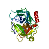


 PDBj
PDBj


 Assembly
 Type: peptide-like
 Mass: 18.015 Da / Num. of mol.: 145 / Source method: isolated from a natural source / Formula: H2O
 Sample preparation
 Processing