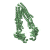
 Movie
Movie Controller


+ Open data

- Basic information
| Entry | Database: PDB / ID: 1cg2 | ||||||
|---|---|---|---|---|---|---|---|
| Title | CARBOXYPEPTIDASE G2 | ||||||
 Components Components | CARBOXYPEPTIDASE G2 Glutamate carboxypeptidase Glutamate carboxypeptidase | ||||||
 Keywords Keywords | METALLOCARBOXYPEPTIDASE / HYDROLASE | ||||||
| Function / homology |  Function and homology informationglutamate carboxypeptidase / carboxypeptidase activity / metallopeptidase activity / proteolysis / metal ion binding Function and homology informationglutamate carboxypeptidase / carboxypeptidase activity / metallopeptidase activity / proteolysis / metal ion bindingSimilarity search - Function | ||||||
| Biological species |  Pseudomonas sp. (bacteria) Pseudomonas sp. (bacteria) | ||||||
| Method | X-RAY DIFFRACTION / SYNCHROTRON / MIR / Resolution: 2.5 Å | ||||||
 Authors Authors | Rowsell, S. / Pauptit, R.A. / Tucker, A.D. / Melton, R.G. / Blow, D.M. / Brick, P. | ||||||
 Citation Citation | Journal: Structure / Year: 1997 Title: Crystal structure of carboxypeptidase G2, a bacterial enzyme with applications in cancer therapy. Authors: Rowsell, S. / Pauptit, R.A. / Tucker, A.D. / Melton, R.G. / Blow, D.M. / Brick, P. #1: Journal: Acta Crystallogr.,Sect.D / Year: 1996Title: A New Crystal Form of Carboxypeptidase G2 from Pseudomonas Sp. Strain Rs-16 which is More Amenable to Structure Determination Authors: Tucker, A.D. / Rowsell, S. / Melton, R.G. / Pauptit, R.A. | ||||||
| History |
|
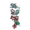
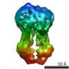
- Structure visualization
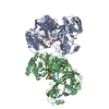
| Structure viewer | Molecule: MolmilJmol/JSmol |
|---|
- Downloads & links
-Download
| PDBx/mmCIF format | 1cg2.cif.gz | 290.6 KB | Display | PDBx/mmCIF format |
|---|---|---|---|---|
| PDB format | pdb1cg2.ent.gz | 231.5 KB | Display | PDB format |
| PDBx/mmJSON format | 1cg2.json.gz | Tree view | PDBx/mmJSON format | |
| Others |  Other downloads Other downloads |
-Validation report
| Arichive directory | https://data.pdbj.org/pub/pdb/validation_reports/cg/1cg2ftp://data.pdbj.org/pub/pdb/validation_reports/cg/1cg2 | HTTPS FTP |
|---|
-Related structure data
| Similar structure data |
|---|

-Links
 PDBj
PDBj
- Assembly
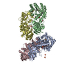
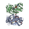
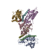
| Deposited unit | 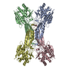
| ||||||||
|---|---|---|---|---|---|---|---|---|---|
| 1 |
| ||||||||
| Unit cell |
| ||||||||
| Details | THERE ARE TWO MOLECULAR DIMERS CONTAINED WITHIN THE CRYSTALLOGRAPHIC ASYMMETRIC UNIT. THE FOUR SUBUNITS PACK TOGETHER WITH APPROXIMATE D2 SYMMETRY. THE SUBUNITS OF ONE MOLECULE HAVE BEEN ASSIGNED CHAIN IDENTIFIERS *A* AND *D*, WHILE THE SUBUNITS OF THE OTHER MOLECULE HAVE BEEN ASSIGNED CHAIN IDENTIFIERS *B* AND *C*. |
-Components
| #1: Protein | Glutamate carboxypeptidase Mass: 41748.359 Da / Num. of mol.: 4 Source method: isolated from a genetically manipulated source Source: (gene. exp.) Pseudomonas sp. (bacteria) / Strain: RS-16 / Cell line: 293 / Production host: Escherichia coli (E. coli) / Strain (production host): 293 / References: UniProt: P06621, glutamate carboxypeptidase#2: Chemical | ChemComp-ZN /   Mass: 65.409 Da / Num. of mol.: 13 / Source method: obtained synthetically / Formula: Zn Mass: 65.409 Da / Num. of mol.: 13 / Source method: obtained synthetically / Formula: Zn#3: Water | ChemComp-HOH / | Water Mass: 18.015 Da / Num. of mol.: 331 / Source method: isolated from a natural source / Formula: H2O Mass: 18.015 Da / Num. of mol.: 331 / Source method: isolated from a natural source / Formula: H2OCompound details | EACH MONOMER CONSISTS OF TWO SEPARATE DOMAINS: A CATALYTIC DOMAIN CONTAINING RESIDUES 26 - 213, 326 ...EACH MONOMER CONSISTS OF TWO SEPARATE DOMAINS: A CATALYTIC DOMAIN CONTAINING | |
|---|
-Experimental details
-Experiment
| Experiment | Method: X-RAY DIFFRACTION / Number of used crystals: 12 |
|---|
- Sample preparation
| Crystal | Density Matthews: 2.9 Å3/Da / Density % sol: 58 % | ||||||||||||||||||||||||||||||||||||||||||||||||||
|---|---|---|---|---|---|---|---|---|---|---|---|---|---|---|---|---|---|---|---|---|---|---|---|---|---|---|---|---|---|---|---|---|---|---|---|---|---|---|---|---|---|---|---|---|---|---|---|---|---|---|---|
| Crystal grow | Temperature: 291 K / Method: vapor diffusion, hanging drop / pH: 7.2 Details: HANGING DROPS WERE FORMED BY MIXING 4 MICROLITERS OF PROTEIN SOLUTION AT 16-20 MG/ML WITH 4 MICROLITERS OF RESERVOIR SOLUTION CONTAINING 10-12% PEG 4000, 0.2M TRIS (PH 7.2), 0.2M ZINC ...Details: HANGING DROPS WERE FORMED BY MIXING 4 MICROLITERS OF PROTEIN SOLUTION AT 16-20 MG/ML WITH 4 MICROLITERS OF RESERVOIR SOLUTION CONTAINING 10-12% PEG 4000, 0.2M TRIS (PH 7.2), 0.2M ZINC ACETATE 10% GLYCEROL. CRYSTALS GREW AT 18-20 DEGREES WITHIN A FEW DAYS., vapor diffusion - hanging drop, temperature 291K | ||||||||||||||||||||||||||||||||||||||||||||||||||
| Crystal grow | *PLUS Method: vapor diffusion, hanging drop | ||||||||||||||||||||||||||||||||||||||||||||||||||
| Components of the solutions | *PLUS
|
-Data collection
| Diffraction | Mean temperature: 293 K |
|---|---|
| Diffraction source | Source: SYNCHROTRON / Site: SRS  / Beamline: PX9.5 / Wavelength: 0.92 / Beamline: PX9.5 / Wavelength: 0.92 |
| Detector | Type: MARRESEARCH / Detector: IMAGE PLATE / Date: Apr 18, 1996 |
| Radiation | Monochromator: Y / Monochromatic (M) / Laue (L): M / Scattering type: x-ray |
| Radiation wavelength | Wavelength: 0.92 Å / Relative weight: 1 |
| Reflection | Resolution: 2.5→20 Å / Num. obs: 62936 / % possible obs: 93.9 % / Observed criterion σ(I): 0 / Redundancy: 3.57 % / Biso Wilson estimate: 41.2 Å2 / Rmerge(I) obs: 0.069 / Rsym value: 0.069 / Net I/σ(I): 6.41 |
| Reflection shell | Resolution: 2.5→2.63 Å / Redundancy: 3.3 % / Rmerge(I) obs: 0.168 / Mean I/σ(I) obs: 4 / % possible all: 90.4 |
| Reflection | *PLUS Num. measured all: 224872 |
| Reflection shell | *PLUS % possible obs: 90.4 % |
- Processing
| Software |
| ||||||||||||||||||||||||||||||||||||||||||||||||||||||||||||||||||||||||||||||||
|---|---|---|---|---|---|---|---|---|---|---|---|---|---|---|---|---|---|---|---|---|---|---|---|---|---|---|---|---|---|---|---|---|---|---|---|---|---|---|---|---|---|---|---|---|---|---|---|---|---|---|---|---|---|---|---|---|---|---|---|---|---|---|---|---|---|---|---|---|---|---|---|---|---|---|---|---|---|---|---|---|---|
| Refinement | Method to determine structure: MIR / Resolution: 2.5→20 Å / Rfactor Rfree error: 0.0045 / Data cutoff high absF: 0 / Data cutoff low absF: 0 / Isotropic thermal model: RESTRAINED / Cross valid method: THROUGHOUT / σ(F): 0
| ||||||||||||||||||||||||||||||||||||||||||||||||||||||||||||||||||||||||||||||||
| Displacement parameters | Biso mean: 37.5 Å2 | ||||||||||||||||||||||||||||||||||||||||||||||||||||||||||||||||||||||||||||||||
| Refinement step | Cycle: LAST / Resolution: 2.5→20 Å
| ||||||||||||||||||||||||||||||||||||||||||||||||||||||||||||||||||||||||||||||||
| Refine LS restraints |
| ||||||||||||||||||||||||||||||||||||||||||||||||||||||||||||||||||||||||||||||||
| Refine LS restraints NCS | NCS model details: RESTRAINTS / Rms dev Biso : 3 Å2 / Weight position: 100 | ||||||||||||||||||||||||||||||||||||||||||||||||||||||||||||||||||||||||||||||||
| LS refinement shell | Resolution: 2.5→2.61 Å / Rfactor Rfree error: 0.018 / Total num. of bins used: 8
| ||||||||||||||||||||||||||||||||||||||||||||||||||||||||||||||||||||||||||||||||
| Xplor file |
| ||||||||||||||||||||||||||||||||||||||||||||||||||||||||||||||||||||||||||||||||
| Software | *PLUS Name: X-PLOR / Version: 3.1 / Classification: refinement | ||||||||||||||||||||||||||||||||||||||||||||||||||||||||||||||||||||||||||||||||
| Refinement | *PLUS | ||||||||||||||||||||||||||||||||||||||||||||||||||||||||||||||||||||||||||||||||
| Solvent computation | *PLUS | ||||||||||||||||||||||||||||||||||||||||||||||||||||||||||||||||||||||||||||||||
| Displacement parameters | *PLUS | ||||||||||||||||||||||||||||||||||||||||||||||||||||||||||||||||||||||||||||||||
| Refine LS restraints | *PLUS
| ||||||||||||||||||||||||||||||||||||||||||||||||||||||||||||||||||||||||||||||||
| LS refinement shell | *PLUS Rfactor obs: 0.196 |
