 Movie
Movie Controller

[English] 日本語
 Yorodumi
- PDB-1c8t: HUMAN STROMELYSIN-1 (E202Q) CATALYTIC DOMAIN COMPLEXED WITH RO-26-2812 -
+ Open data

- Basic information
| Entry | Database: PDB / ID: 1c8t | ||||||
|---|---|---|---|---|---|---|---|
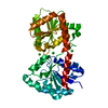
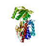
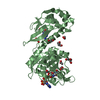
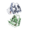
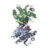
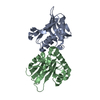
| Title | HUMAN STROMELYSIN-1 (E202Q) CATALYTIC DOMAIN COMPLEXED WITH RO-26-2812 | ||||||
 Components Components | STROMELYSIN-1 Matrix metalloproteinase Matrix metalloproteinase | ||||||
 Keywords Keywords | HYDROLASE/HYDROLASE INHIBITOR / PROTEIN-INHIBITOR COMPLEX / MUTANT PROTEIN / HYDROLASE-HYDROLASE INHIBITOR COMPLEX | ||||||
| Function / homology |  Function and homology informationstromelysin 1 / cellular response to UV-A / regulation of neuroinflammatory response / Assembly of collagen fibrils and other multimeric structures / Activation of Matrix Metalloproteinases / response to amyloid-beta / Collagen degradation / collagen catabolic process / extracellular matrix disassembly / cellular response to nitric oxide ...stromelysin 1 / cellular response to UV-A / regulation of neuroinflammatory response / Assembly of collagen fibrils and other multimeric structures / Activation of Matrix Metalloproteinases / response to amyloid-beta / Collagen degradation / collagen catabolic process / extracellular matrix disassembly / cellular response to nitric oxide / negative regulation of reactive oxygen species metabolic process / EGFR Transactivation by Gastrin / extracellular matrix / Degradation of the extracellular matrix / extracellular matrix organization / positive regulation of protein-containing complex assembly / metalloendopeptidase activity / cellular response to reactive oxygen species / metallopeptidase activity / peptidase activity / Interleukin-4 and Interleukin-13 signaling / endopeptidase activity / cellular response to lipopolysaccharide / Extra-nuclear estrogen signaling / serine-type endopeptidase activity / innate immune response / proteolysis / extracellular space / zinc ion binding / extracellular region / nucleus / cytoplasm Function and homology informationstromelysin 1 / cellular response to UV-A / regulation of neuroinflammatory response / Assembly of collagen fibrils and other multimeric structures / Activation of Matrix Metalloproteinases / response to amyloid-beta / Collagen degradation / collagen catabolic process / extracellular matrix disassembly / cellular response to nitric oxide ...stromelysin 1 / cellular response to UV-A / regulation of neuroinflammatory response / Assembly of collagen fibrils and other multimeric structures / Activation of Matrix Metalloproteinases / response to amyloid-beta / Collagen degradation / collagen catabolic process / extracellular matrix disassembly / cellular response to nitric oxide / negative regulation of reactive oxygen species metabolic process / EGFR Transactivation by Gastrin / extracellular matrix / Degradation of the extracellular matrix / extracellular matrix organization / positive regulation of protein-containing complex assembly / metalloendopeptidase activity / cellular response to reactive oxygen species / metallopeptidase activity / peptidase activity / Interleukin-4 and Interleukin-13 signaling / endopeptidase activity / cellular response to lipopolysaccharide / Extra-nuclear estrogen signaling / serine-type endopeptidase activity / innate immune response / proteolysis / extracellular space / zinc ion binding / extracellular region / nucleus / cytoplasmSimilarity search - Function | ||||||
| Biological species |  Homo sapiens (human) Homo sapiens (human) | ||||||
| Method | X-RAY DIFFRACTION / Resolution: 2.6 Å | ||||||
 Authors Authors | Steele, D.L. / el-Kabbani, O. / Dunten, P. / Crowther, R.L. | ||||||
 Citation Citation | Journal: Protein Eng. / Year: 2000 Title: Expression, characterization and structure determination of an active site mutant (Glu202-Gln) of mini-stromelysin-1. Authors: Steele, D.L. / El-Kabbani, O. / Dunten, P. / Windsor, L.J. / Kammlott, R.U. / Crowther, R.L. / Michoud, C. / Engler, J.A. / Birktoft, J.J. | ||||||
| History |
|
- Structure visualization
| Structure viewer | Molecule: MolmilJmol/JSmol |
|---|
- Downloads & links
-Download
| PDBx/mmCIF format | 1c8t.cif.gz | 83.1 KB | Display | PDBx/mmCIF format |
|---|---|---|---|---|
| PDB format | pdb1c8t.ent.gz | 62.1 KB | Display | PDB format |
| PDBx/mmJSON format | 1c8t.json.gz | Tree view | PDBx/mmJSON format | |
| Others |  Other downloads Other downloads |
-Validation report
| Arichive directory | https://data.pdbj.org/pub/pdb/validation_reports/c8/1c8tftp://data.pdbj.org/pub/pdb/validation_reports/c8/1c8t | HTTPS FTP |
|---|
-Related structure data


-Links
 PDBj
PDBj


- Assembly
| Deposited unit | 
| ||||||||
|---|---|---|---|---|---|---|---|---|---|
| 1 |
| ||||||||
| Unit cell |
|
-Components
| #1: Protein | Matrix metalloproteinase Mass: 18692.777 Da / Num. of mol.: 2 / Fragment: CATALYTIC DOMAIN / Mutation: E202Q, S252P Source method: isolated from a genetically manipulated source Source: (gene. exp.) Homo sapiens (human) / Cell line: GIGIVAL FIBROBLASTS / Plasmid: PGEMEX-2 / Production host:  Escherichia coli (E. coli) / References: UniProt: P08254, stromelysin 1 Escherichia coli (E. coli) / References: UniProt: P08254, stromelysin 1#2: Chemical | ChemComp-ZN /   Mass: 65.409 Da / Num. of mol.: 4 / Source method: obtained synthetically / Formula: Zn / Details: Synthetic compound Mass: 65.409 Da / Num. of mol.: 4 / Source method: obtained synthetically / Formula: Zn / Details: Synthetic compound#3: Chemical | ChemComp-CA /   Mass: 40.078 Da / Num. of mol.: 6 / Source method: obtained synthetically / Formula: Ca Mass: 40.078 Da / Num. of mol.: 6 / Source method: obtained synthetically / Formula: Ca#4: Chemical |   Mass: 457.586 Da / Num. of mol.: 2 / Source method: obtained synthetically / Formula: C24H31N3O4S / Details: SYNTHETIC INHIBITOR BASED ON PRO-PEPTIDE SEQUENCE Mass: 457.586 Da / Num. of mol.: 2 / Source method: obtained synthetically / Formula: C24H31N3O4S / Details: SYNTHETIC INHIBITOR BASED ON PRO-PEPTIDE SEQUENCE#5: Water | ChemComp-HOH / | Water Mass: 18.015 Da / Num. of mol.: 71 / Source method: isolated from a natural source / Formula: H2O Mass: 18.015 Da / Num. of mol.: 71 / Source method: isolated from a natural source / Formula: H2O |
|---|
-Experimental details
-Experiment
| Experiment | Method: X-RAY DIFFRACTION / Number of used crystals: 2 |
|---|
- Sample preparation
| Crystal | Density Matthews: 2.07 Å3/Da / Density % sol: 40.6 % | ||||||||||||||||||||||||||||
|---|---|---|---|---|---|---|---|---|---|---|---|---|---|---|---|---|---|---|---|---|---|---|---|---|---|---|---|---|---|
| Crystal grow | Temperature: 295 K / Method: vapor diffusion, hanging drop / pH: 7 Details: PEG, potassium chloride, calcium chloride, cacodylate, pH 7, VAPOR DIFFUSION, HANGING DROP, temperature 295K | ||||||||||||||||||||||||||||
| Crystal grow | *PLUS Temperature: 22 ℃ / PH range low: 7 / PH range high: 6.5 | ||||||||||||||||||||||||||||
| Components of the solutions | *PLUS
|
-Data collection
| Diffraction | Mean temperature: 100 K |
|---|---|
| Diffraction source | Source: ROTATING ANODE / Type: RIGAKU RU200 / Wavelength: 1.5418 |
| Detector | Type: SDMS / Detector: AREA DETECTOR / Date: Jan 1, 1995 |
| Radiation | Protocol: SINGLE WAVELENGTH / Monochromatic (M) / Laue (L): M / Scattering type: x-ray |
| Radiation wavelength | Wavelength: 1.5418 Å / Relative weight: 1 |
| Reflection | Highest resolution: 2.15 Å / Num. all: 17584 / Num. obs: 15629 / Redundancy: 5.9 % / Rmerge(I) obs: 0.079 / Net I/σ(I): 19 |
| Reflection shell | Highest resolution: 2.15 Å |
| Reflection | *PLUS % possible obs: 88.9 % |
- Processing
| Software |
| ||||||||||||
|---|---|---|---|---|---|---|---|---|---|---|---|---|---|
| Refinement | Resolution: 2.6→20 Å /
| ||||||||||||
| Refinement step | Cycle: LAST / Resolution: 2.6→20 Å
| ||||||||||||
| Software | *PLUS Name: REFMAC / Classification: refinement | ||||||||||||
| Refine LS restraints | *PLUS
|
