 Movie
Movie Controller

[English] 日本語
 Yorodumi
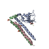
- PDB-1c7u: Complex of the DNA binding core domain of the transcription facto... -
+ Open data

- Basic information
| Entry | Database: PDB / ID: 1c7u | ||||||
|---|---|---|---|---|---|---|---|
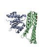
| Title | Complex of the DNA binding core domain of the transcription factor MEF2A with a 20mer oligonucleotide | ||||||
 Components Components |
| ||||||
 Keywords Keywords | TRANSCRIPTION/DNA /  DNA BINDING PROTEIN / TRANSCRIPTION FACTOR / MADS-BOX / SAM DOMAIN / TRANSCRIPTION-DNA COMPLEX DNA BINDING PROTEIN / TRANSCRIPTION FACTOR / MADS-BOX / SAM DOMAIN / TRANSCRIPTION-DNA COMPLEX | ||||||
| Function / homology |  Function and homology information Function and homology informationERK5 cascade / ventricular cardiac myofibril assembly / mitochondrion distribution / cardiac conduction / mitochondrial genome maintenance / dendrite morphogenesis / muscle organ development / histone acetyltransferase binding / Myogenesis / positive regulation of cardiac muscle hypertrophy ...ERK5 cascade / ventricular cardiac myofibril assembly / mitochondrion distribution / cardiac conduction / mitochondrial genome maintenance / dendrite morphogenesis / muscle organ development / histone acetyltransferase binding / Myogenesis / positive regulation of cardiac muscle hypertrophy / SMAD binding / ERK/MAPK targets / cellular response to calcium ion / positive regulation of glucose import / RNA polymerase II transcription regulatory region sequence-specific DNA binding / histone deacetylase binding / MAPK cascade / heart development / DNA-binding transcription activator activity, RNA polymerase II-specific / DNA-binding transcription factor binding / transcription regulator complex / RNA polymerase II-specific DNA-binding transcription factor binding / sequence-specific DNA binding / cell differentiation / DNA-binding transcription factor activity, RNA polymerase II-specific / RNA polymerase II cis-regulatory region sequence-specific DNA binding / DNA-binding transcription factor activity / protein heterodimerization activity / DNA-templated transcription / apoptotic process / chromatin binding / chromatin / positive regulation of gene expression / protein kinase binding / negative regulation of transcription by RNA polymerase II / positive regulation of transcription by RNA polymerase II / nucleoplasm / nucleus / cytosolSimilarity search - Function | ||||||
| Biological species |  Homo sapiens (human) Homo sapiens (human) | ||||||
| Method | SOLUTION NMR / simulated annealing | ||||||
 Authors Authors | Clore, G.M. / Huang, K. | ||||||
 Citation Citation | Journal: Embo J. / Year: 2000 Title: Solution structure of the MEF2A-DNA complex: structural basis for the modulation of DNA bending and specificity by MADS-box transcription factors Authors: Huang, K. / Louis, J.M. / Donaldson, L. / Lim, F.L. / Sharrocks, A.D. / Clore, G.M. | ||||||
| History |
|
- Structure visualization
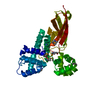
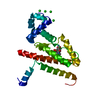
| Structure viewer | Molecule: MolmilJmol/JSmol |
|---|
- Downloads & links
-Download
| PDBx/mmCIF format | 1c7u.cif.gz | 98 KB | Display | PDBx/mmCIF format |
|---|---|---|---|---|
| PDB format | pdb1c7u.ent.gz | 72.8 KB | Display | PDB format |
| PDBx/mmJSON format | 1c7u.json.gz | Tree view | PDBx/mmJSON format | |
| Others |  Other downloads Other downloads |
-Validation report
| Arichive directory | https://data.pdbj.org/pub/pdb/validation_reports/c7/1c7uftp://data.pdbj.org/pub/pdb/validation_reports/c7/1c7u | HTTPS FTP |
|---|
-Related structure data
| Similar structure data |
|---|


-Links
 PDBj
PDBj


- Assembly
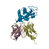
| Deposited unit | 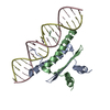
| |||||||||
|---|---|---|---|---|---|---|---|---|---|---|
| 1 |
| |||||||||
| NMR ensembles |
|
-Components
| #1: DNA chain | Mass: 6133.979 Da / Num. of mol.: 2 / Source method: obtained synthetically #2: Protein | Mass: 10007.484 Da / Num. of mol.: 2 / Fragment: RESIDUES 2-86 Source method: isolated from a genetically manipulated source Source: (gene. exp.) Homo sapiens (human) / Plasmid: PET11A / Production host:  Escherichia coli (E. coli) / Strain (production host): BE23 / References: UniProt: Q02078 Escherichia coli (E. coli) / Strain (production host): BE23 / References: UniProt: Q02078 |
|---|
-Experimental details
-Experiment
| Experiment | Method: SOLUTION NMR |
|---|---|
| NMR experiment | Type: (1) TRIPLE RESONANCE FOR ASSIGNMENT OF PROTEIN. (2) QUANTITATIVE J CORRELATION FOR COUPLING CONSTANTS. (3) 3D AND 4D HETERONUCLEAR SEPARATED AND FILTERED NOE EXPTS. (4) 2D 12C-FILTERED ...Type: (1) TRIPLE RESONANCE FOR ASSIGNMENT OF PROTEIN. (2) QUANTITATIVE J CORRELATION FOR COUPLING CONSTANTS. (3) 3D AND 4D HETERONUCLEAR SEPARATED AND FILTERED NOE EXPTS. (4) 2D 12C-FILTERED EXPERIMENTS FOR DNA ASSIGNMENTS. (5) IPAP EXPTS FOR DIPOLAR COUPLINGS DIPOLAR COUPLINGS WERE MEASURED IN A BICELLE LIQUID CRYSTALLINE MEDIUM. |
| NMR details | Text: SOLVED BY MULTI HETERONUCLEAR NMR AND IS BASED ON 4560 EXPERIMENTAL NMR RESTRAINTS (I.E. 2280 UNIQUE ONES SINCE PROTEIN IS A HOMODIMER AND DNA IS PALINDROMIC). NOE RESTRAINTS: (A) PROTEIN: 664 ...Text: SOLVED BY MULTI HETERONUCLEAR NMR AND IS BASED ON 4560 EXPERIMENTAL NMR RESTRAINTS (I.E. 2280 UNIQUE ONES SINCE PROTEIN IS A HOMODIMER AND DNA IS PALINDROMIC). NOE RESTRAINTS: (A) PROTEIN: 664 SEQUENTIAL, 504 MEDIUM RANGE, 212 LONG RANGE, 616 INTRARESIDUE, 174 INTERSUBUNIT. (B) DNA: 428 INTRARESIDUE, 196 SEQUENTIAL INTRASTRAND, 24 INTERSTRAND. (C) PROTEIN- DNA 168. H-BOND RESTRAINTS: PROTEIN 138, DNA 120. TORSION ANGLE RESTRAINTS: PROTEIN 480 (142 PHI, 142 PSI, 112 CHI1, 68 CHI2, 16 CHI3), DNA 228. THREE-BOND HN-HALPHA COUPLING CONSTANTS: 72. SECONDARY 13C SHIFTS: 140 13CALPHA, 140 13CBETA. DIPOLAR COUPLINGS: 1DNH PROTEIN: 70, 1DCH DNA 70. REPULSIVE RESTRAINTS: 106 |
- Sample preparation
| Sample conditions | pH: 6.6 / Temperature: 308 K |
|---|---|
| Crystal grow | *PLUS Method: other / Details: NMR |
-NMR measurement
| NMR spectrometer |
|
|---|
- Processing
| NMR software |
| |||||||||
|---|---|---|---|---|---|---|---|---|---|---|
| Refinement | Method: simulated annealing / Software ordinal: 1 Details: THE STRUCTURES WERE CALCULATED USING THE SIMULATED ANNEALING PROTOCOL OF NILGES ET AL. (1988) FEBS LETT. 229, 129-136 USING THE PROGRAM XPLOR/CNS MODIFIED TO INCORPORATE COUPLING CONSTANT ...Details: THE STRUCTURES WERE CALCULATED USING THE SIMULATED ANNEALING PROTOCOL OF NILGES ET AL. (1988) FEBS LETT. 229, 129-136 USING THE PROGRAM XPLOR/CNS MODIFIED TO INCORPORATE COUPLING CONSTANT RESTRAINTS (GARRETT ET AL. (1984) J. MAGN. RESON. SERIES B 104, 99-103), CARBON CHEMICAL SHIFT RESTRAINTS, (KUSZEWSKI ET AL. (1995) J. MAGN. RESON. SERIES B 106, 92-96) RESTRAINTS, RESIDUAL DIPOLAR COUPLING RESTRAINTS (CLORE ET AL. J. MAGN. RESON 131, 159-162 (1998); J. MAGN 133, 216-221 (1998)), AND A CONFORMATIONAL DATABASE POTENTIAL FOR PROTEINS AND NUCLEIC ACIDS (KUSZEWSKI ET AL. PROTEIN SCI. 5, 1067-1080 (1996); J. MAGN. RESON 125, 171-177 (1997)). IN THIS ENTRY THE LAST NUMERICAL COLUMN IS THE RMS OF THE 35 INDIVIDUAL SIMULATED ANNEALING STRUCTURES ABOUT THE MEAN COORDINATE POSITIONS. THE LATTER ARE OBTAINED BY TAKING THE AVERAGE OF THE 35 SIMULATED ANNEALING STRUCTURES BEST-FITTED TO RESIDUES 1-73 AND 101-173 OF THE PROTEIN AND RESIDUES 201-240 OF THE DNA. RESIDUES 74-85 AND 174-185 ARE DISORDERED IN SOLUTION AND ARE THEREFORE NOT INCLUDED IN THE COORDINATES. THE RESTRAINED MINIMIZED MEAN STRUCTURE IS OBTAINED BY RESTRAINED REGULARIZATION OF THE AVERAGE COORDINATES AGAINST THE SAME TARGET FUNCTION USED TO CALCULATE THE SIMULATED ANNEALING STRUCTURES. | |||||||||
| NMR ensemble | Conformer selection criteria: REGULARIZED MEAN STRUCTURE / Conformers calculated total number: 35 / Conformers submitted total number: 1 |
