 Movie
Movie Controller

[English] 日本語
 Yorodumi
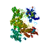
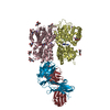
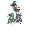
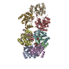
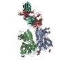
- PDB-1c7s: BETA-N-ACETYLHEXOSAMINIDASE MUTANT D539A COMPLEXED WITH DI-N-ACET... -
+ Open data

- Basic information
| Entry | Database: PDB / ID: 1c7s | |||||||||
|---|---|---|---|---|---|---|---|---|---|---|
| Title | BETA-N-ACETYLHEXOSAMINIDASE MUTANT D539A COMPLEXED WITH DI-N-ACETYL-BETA-D-GLUCOSAMINE (CHITOBIASE) | |||||||||
 Components Components | BETA-N-ACETYLHEXOSAMINIDASE Hexosaminidase Hexosaminidase | |||||||||
 Keywords Keywords | HYDROLASE / GLYCOSYL HYDROLASE / BETA-N-ACETYLHEXOSAMINIDASE / CHITINOLYSIS / A/B(TIM)-BARREL / SITE DIRECTED MUTAGENESIS / SUBSTRATE NUCLEOPHILE STABILIZER MUTATION | |||||||||
| Function / homology |  Function and homology informationbeta-N-acetylhexosaminidase / beta-N-acetylhexosaminidase activity / N-acetyl-beta-D-galactosaminidase activity / chitin catabolic process / polysaccharide binding / polysaccharide catabolic process / periplasmic space Function and homology informationbeta-N-acetylhexosaminidase / beta-N-acetylhexosaminidase activity / N-acetyl-beta-D-galactosaminidase activity / chitin catabolic process / polysaccharide binding / polysaccharide catabolic process / periplasmic spaceSimilarity search - Function | |||||||||
| Biological species |  Serratia marcescens (bacteria) Serratia marcescens (bacteria) | |||||||||
| Method | X-RAY DIFFRACTION / SYNCHROTRON / MOLECULAR REPLACEMENT / Resolution: 1.8 Å | |||||||||
 Authors Authors | Prag, G. / Papanikolau, Y. / Tavlas, G. / Vorgias, C.E. / Petratos, K. / Oppenheim, A.B. | |||||||||
 Citation Citation | Journal: J.Mol.Biol. / Year: 2000 Title: Structures of chitobiase mutants complexed with the substrate Di-N-acetyl-d-glucosamine: the catalytic role of the conserved acidic pair, aspartate 539 and glutamate 540. Authors: Prag, G. / Papanikolau, Y. / Tavlas, G. / Vorgias, C.E. / Petratos, K. / Oppenheim, A.B. #1: Journal: Nat.Struct.Biol. / Year: 1996Title: Bacterial Chitobiase Structure Provides Insight Into Catalytic Mechanism and the Basis of Tay-Sachs Disease Authors: Tews, I. / Perrakis, A. / Oppenheim, A. / Dauter, Z. / Wilson, K.S. / Vorgias, C.E. | |||||||||
| History |
|
- Structure visualization
| Structure viewer | Molecule: MolmilJmol/JSmol |
|---|
- Downloads & links
-Download
| PDBx/mmCIF format | 1c7s.cif.gz | 203.4 KB | Display | PDBx/mmCIF format |
|---|---|---|---|---|
| PDB format | pdb1c7s.ent.gz | 155.9 KB | Display | PDB format |
| PDBx/mmJSON format | 1c7s.json.gz | Tree view | PDBx/mmJSON format | |
| Others |  Other downloads Other downloads |
-Validation report
| Arichive directory | https://data.pdbj.org/pub/pdb/validation_reports/c7/1c7sftp://data.pdbj.org/pub/pdb/validation_reports/c7/1c7s | HTTPS FTP |
|---|
-Related structure data
| Related structure data |  1c7tC 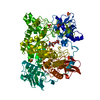 1qbbS S: Starting model for refinement C: citing same article ( |
|---|---|
| Similar structure data |


-Links
 PDBj
PDBj
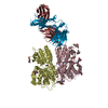
- Assembly
| Deposited unit | 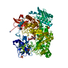
| ||||||||
|---|---|---|---|---|---|---|---|---|---|
| 1 | 
| ||||||||
| Unit cell |
|
-Components
| #1: Protein | Hexosaminidase / N-ACETYL-BETA-D-GLUCOSAMINIDASE / CHITOBIASE Mass: 95940.930 Da / Num. of mol.: 1 Fragment: MATURE PROTEIN, PERIPLASMATIC TARGETING SEQUENCE RESIDUES 1-27 CLEAVED OFF DURING MATURATION Mutation: D539A Source method: isolated from a genetically manipulated source Details: COMPLEXED WITH DINAG / Source: (gene. exp.) Serratia marcescens (bacteria) / Strain: A9301 / Plasmid: PKK177-3 / Cellular location (production host): PERIPLASM / Production host: Escherichia coli (E. coli) / Strain (production host): XL1-BLUE / References: UniProt: Q54468, beta-N-acetylhexosaminidase | ||
|---|---|---|---|
| #2: Polysaccharide | 2-acetamido-2-deoxy-beta-D-glucopyranose-(1-4)-2-acetamido-2-deoxy-beta-D-glucopyranose / Mass: 424.401 Da / Num. of mol.: 1 Source method: isolated from a genetically manipulated source | ||
| #3: Chemical | ChemComp-SO4 / Sulfate  Mass: 96.063 Da / Num. of mol.: 4 / Source method: obtained synthetically / Formula: SO4 Mass: 96.063 Da / Num. of mol.: 4 / Source method: obtained synthetically / Formula: SO4#4: Water | ChemComp-HOH / | Water Mass: 18.015 Da / Num. of mol.: 821 / Source method: isolated from a natural source / Formula: H2O Mass: 18.015 Da / Num. of mol.: 821 / Source method: isolated from a natural source / Formula: H2O |
-Experimental details
-Experiment
| Experiment | Method: X-RAY DIFFRACTION / Number of used crystals: 1 |
|---|
- Sample preparation
| Crystal | Density Matthews: 2.56 Å3/Da / Density % sol: 51.9 % | |||||||||||||||||||||||||
|---|---|---|---|---|---|---|---|---|---|---|---|---|---|---|---|---|---|---|---|---|---|---|---|---|---|---|
| Crystal grow | Method: vapor diffusion, hanging drop / pH: 4.8 Details: CO-CRYSTALS WERE GROWN BY THE HANGING-DROP VAPOR DIFFUSION METHOD. RESERVOIR BUFFER CONTAINED 2.3 MOLAR AMMONIUM SULFATE AND 100 MILLIMOLAR CACODYLATE BUFFER PH 4.8. PROTEIN SOLUTION 40 ...Details: CO-CRYSTALS WERE GROWN BY THE HANGING-DROP VAPOR DIFFUSION METHOD. RESERVOIR BUFFER CONTAINED 2.3 MOLAR AMMONIUM SULFATE AND 100 MILLIMOLAR CACODYLATE BUFFER PH 4.8. PROTEIN SOLUTION 40 MILLIGRAM PER MILLILITER WAS MIXED WITH AN EQUAL VOLUME OF RESERVOIR CONTAINING 10 MILLIMOLAR DI-NAG. CRYSTALS ABOUT 0.5 X 0.2 X 0.2 MILLIMETER IN SIZE WERE FORMED WITHIN 2-3 DAYS., VAPOR DIFFUSION, HANGING DROP | |||||||||||||||||||||||||
| Crystal grow | *PLUS | |||||||||||||||||||||||||
| Components of the solutions | *PLUS
|
-Data collection
| Diffraction | Mean temperature: 100 K |
|---|---|
| Diffraction source | Source: SYNCHROTRON / Site: EMBL/DESY, HAMBURG  / Beamline: X11 / Wavelength: 0.9116 / Beamline: X11 / Wavelength: 0.9116 |
| Detector | Type: MARRESEARCH / Detector: IMAGE PLATE / Date: Apr 4, 1999 / Details: 345 MM IMAGE PLATE |
| Radiation | Protocol: SINGLE WAVELENGTH / Monochromatic (M) / Laue (L): M / Scattering type: x-ray |
| Radiation wavelength | Wavelength: 0.9116 Å / Relative weight: 1 |
| Reflection | Resolution: 1.8→15 Å / Num. obs: 76047 / % possible obs: 96.9 % / Rmerge(I) obs: 0.024 / Net I/σ(I): 32832 |
| Reflection shell | Resolution: 1.8→1.86 Å / Rmerge(I) obs: 0.095 / Mean I/σ(I) obs: 4738 / % possible all: 90.8 |
| Reflection | *PLUS % possible obs: 86.9 % / Num. measured all: 937195 |
| Reflection shell | *PLUS Lowest resolution: 1.95 Å / % possible obs: 90.8 % |
- Processing
| Software |
| ||||||||||||||||||||||||||||||||||||||||||||||||||||||||||||||||||||||||||||||||||||
|---|---|---|---|---|---|---|---|---|---|---|---|---|---|---|---|---|---|---|---|---|---|---|---|---|---|---|---|---|---|---|---|---|---|---|---|---|---|---|---|---|---|---|---|---|---|---|---|---|---|---|---|---|---|---|---|---|---|---|---|---|---|---|---|---|---|---|---|---|---|---|---|---|---|---|---|---|---|---|---|---|---|---|---|---|---|
| Refinement | Method to determine structure: MOLECULAR REPLACEMENT Starting model: PDB ENTRY 1QBB Resolution: 1.8→15 Å / SU B: 2.724 / SU ML: 0.087 / Cross valid method: R-FREE / σ(F): 0 / ESU R: 0.152 / ESU R Free: 0.147
| ||||||||||||||||||||||||||||||||||||||||||||||||||||||||||||||||||||||||||||||||||||
| Displacement parameters | Biso mean: 21.8 Å2 | ||||||||||||||||||||||||||||||||||||||||||||||||||||||||||||||||||||||||||||||||||||
| Refinement step | Cycle: LAST / Resolution: 1.8→15 Å
| ||||||||||||||||||||||||||||||||||||||||||||||||||||||||||||||||||||||||||||||||||||
| Refine LS restraints |
| ||||||||||||||||||||||||||||||||||||||||||||||||||||||||||||||||||||||||||||||||||||
| Software | *PLUS Name: 'REFMAC / ARP' / Classification: refinement | ||||||||||||||||||||||||||||||||||||||||||||||||||||||||||||||||||||||||||||||||||||
| Refinement | *PLUS σ(F): 0 / % reflection Rfree: 5 % | ||||||||||||||||||||||||||||||||||||||||||||||||||||||||||||||||||||||||||||||||||||
| Solvent computation | *PLUS | ||||||||||||||||||||||||||||||||||||||||||||||||||||||||||||||||||||||||||||||||||||
| Displacement parameters | *PLUS Biso mean: 21.8 Å2 | ||||||||||||||||||||||||||||||||||||||||||||||||||||||||||||||||||||||||||||||||||||
| Refine LS restraints | *PLUS
|
