 Movie
Movie Controller

[English] 日本語
 Yorodumi
- PDB-1brd: Model for the structure of Bacteriorhodopsin based on high-resolu... -
+ Open data

- Basic information
| Entry | Database: PDB / ID: 1brd | |||||||||
|---|---|---|---|---|---|---|---|---|---|---|
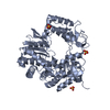
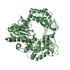
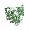
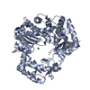
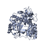
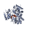
| Title | Model for the structure of Bacteriorhodopsin based on high-resolution Electron Cryo-microscopy | |||||||||
 Components Components | BACTERIORHODOPSIN PRECURSOR | |||||||||
 Keywords Keywords |  PHOTORECEPTOR PHOTORECEPTOR | |||||||||
| Function / homology |  Function and homology informationphotoreceptor activity / phototransduction / proton transmembrane transport / monoatomic ion channel activity / plasma membrane Function and homology informationphotoreceptor activity / phototransduction / proton transmembrane transport / monoatomic ion channel activity / plasma membraneSimilarity search - Function | |||||||||
| Biological species |  Halobacterium salinarum (Halophile) Halobacterium salinarum (Halophile) | |||||||||
| Method | ELECTRON CRYSTALLOGRAPHY / electron crystallography / cryo EM / Resolution: 3.5 Å | |||||||||
 Authors Authors | Henderson, R. / Baldwin, J.M. / Ceska, T.A. / Zemlin, F. / Beckmann, E. / Downing, K.H. | |||||||||
 Citation Citation | Journal: J Mol Biol / Year: 1990 Title: Model for the structure of bacteriorhodopsin based on high-resolution electron cryo-microscopy. Authors: R Henderson / J M Baldwin / T A Ceska / F Zemlin / E Beckmann / K H Downing /  Abstract: The light-driven proton pump bacteriorhodopsin occurs naturally as two-dimensional crystals. A three-dimensional density map of the structure, at near-atomic resolution, has been obtained by studying ...The light-driven proton pump bacteriorhodopsin occurs naturally as two-dimensional crystals. A three-dimensional density map of the structure, at near-atomic resolution, has been obtained by studying the crystals using electron cryo-microscopy to obtain electron diffraction patterns and high-resolution micrographs. New methods were developed for analysing micrographs from tilted specimens, incorporating methods previously developed for untilted specimens that enable large areas to be analysed and corrected for distortions. Data from 72 images, from both tilted and untilted specimens, were analysed to produce the phases of 2700 independent Fourier components of the structure. The amplitudes of these components were accurately measured from 150 diffraction patterns. Together, these data represent about half of the full three-dimensional transform to 3.5 A. The map of the structure has a resolution of 3.5 A in a direction parallel to the membrane plane but lower than this in the perpendicular direction. It shows many features in the density that are resolved from the main density of the seven alpha-helices. We interpret these features as the bulky aromatic side-chains of phenylalanine, tyrosine and tryptophan residues. There is also a very dense feature, which is the beta-ionone ring of the retinal chromophore. Using these bulky side-chains as guide points and taking account of bulges in the helices that indicate smaller side-chains such as leucine, a complete atomic model for bacteriorhodopsin between amino acid residues 8 and 225 has been built. There are 21 amino acid residues, contributed by all seven helices, surrounding the retinal and 26 residues, contributed by five helices, forming the proton pathway or channel. Ten of the amino acid residues in the middle of the proton channel are also part of the retinal binding site. The model also provides a useful basis for consideration of the mechanism of proton pumping and allows a consistent interpretation of a great deal of other experimental data. In particular, the structure suggests that pK changes in the Schiff base must act as the means by which light energy is converted into proton pumping pressure in the channel. Asp96 is on the pathway from the cytoplasm to the Schiff base and Asp85 is on the pathway from the Schiff base to the extracellular surface. #1: Journal: J.Mol.Biol. / Year: 1978Title: Specific Labelling of the Protein and Lipid on the Extracellular Surface of Purple Membrane Authors: Henderson, R. / Jubb, J.S. / Whytock, S. #2: Journal: Nature / Year: 1975Title: Three-Dimensional Model of Purple Membrane Obtained by Electron Microscopy Authors: Henderson, R. / Unwin, P.N.T. | |||||||||
| History |
| |||||||||
| Remark 650 | HELIX THE ENDS OF THE HELICES ARE UNCERTAIN BY AT LEAST ONE RESIDUE. IN ADDITION, PRO 50, PRO 91, ...HELIX THE ENDS OF THE HELICES ARE UNCERTAIN BY AT LEAST ONE RESIDUE. IN ADDITION, PRO 50, PRO 91, AND PRO 186 ARE IN THE MIDDLE OF HELICAL SEGMENTS BUT IN EACH CASE THE HELICES ARE BENT NEAR THE PROLINE RESIDUES. |
- Structure visualization
| Movie |
Movie viewer |
|---|---|
| Structure viewer | Molecule: MolmilJmol/JSmol |
- Downloads & links
-Download
| PDBx/mmCIF format | 1brd.cif.gz | 40.5 KB | Display | PDBx/mmCIF format |
|---|---|---|---|---|
| PDB format | pdb1brd.ent.gz | 28.4 KB | Display | PDB format |
| PDBx/mmJSON format | 1brd.json.gz | Tree view | PDBx/mmJSON format | |
| Others |  Other downloads Other downloads |
-Validation report
| Arichive directory | https://data.pdbj.org/pub/pdb/validation_reports/br/1brdftp://data.pdbj.org/pub/pdb/validation_reports/br/1brd | HTTPS FTP |
|---|
-Related structure data
| Similar structure data |
|---|


-Links
 PDBj
PDBj


- Assembly
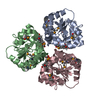
| Deposited unit | 
| ||||||||
|---|---|---|---|---|---|---|---|---|---|
| 1 | 
| ||||||||
| Unit cell |
| ||||||||
| Atom site foot note | 1: A TEMPERATURE FACTOR OF 99.0 DENOTES SIDE CHAINS WITH NO CLEAR DENSITY IN THE EXPERIMENTAL MAP OR WITH OTHER AMBIGUITY. A TEMPERATURE FACTOR OF 20.0 DENOTES GOOD SIDE CHAINS OR THE POLYPEPTIDE ...1: A TEMPERATURE FACTOR OF 99.0 DENOTES SIDE CHAINS WITH NO CLEAR DENSITY IN THE EXPERIMENTAL MAP OR WITH OTHER AMBIGUITY. A TEMPERATURE FACTOR OF 20.0 DENOTES GOOD SIDE CHAINS OR THE POLYPEPTIDE BACKBONE IN THE MAIN HELICAL SEGMENTS. SOME SIDE CHAINS OF INTERMEDIATE QUALITY HAVE TEMPERATURE FACTORS OF 40.0. |
-Components
| #1: Protein | Mass: 26797.381 Da / Num. of mol.: 1 Source method: isolated from a genetically manipulated source Source: (gene. exp.) Halobacterium salinarum (Halophile) / References: UniProt: P02945 |
|---|---|
| #2: Chemical | ChemComp-RET / Retinal  Mass: 284.436 Da / Num. of mol.: 1 / Source method: obtained synthetically / Formula: C20H28O Mass: 284.436 Da / Num. of mol.: 1 / Source method: obtained synthetically / Formula: C20H28O |
-Experimental details
-Experiment
| Experiment | Method: ELECTRON CRYSTALLOGRAPHY |
|---|---|
| EM experiment | Aggregation state: 2D ARRAY / 3D reconstruction method: electron crystallography |
| Crystal symmetry | ∠γ: 120 ° / C sampling length: 100 Å / A: 62.45 Å / B: 62.45 Å / C: 100 Å / Space group name H-M: P3 |
- Sample preparation
| Component | Name: Bacteriorhodopsin from purple membrane / Type: COMPLEX |
|---|---|
| Buffer solution | pH: 5.2 Details: 0.1 mM potassium phosphate, 6 mM octyl glucoside, 0.2 mM trimethylammonium chloride |
| Specimen | Conc.: 3 mg/ml / Embedding applied: YES / Shadowing applied: NO / Staining applied: NO / Vitrification applied: YES |
| Specimen support | Details: carbon aged several days to optimize hydrophobicity Grid material: COPPER |
| EM embedding | Details: 0.8% (w/v) glucose / Material: glucose |
-Data collection
| EM imaging | Specimen-ID: 1
| ||||||||||||||||||||||||||||||||||||||||
|---|---|---|---|---|---|---|---|---|---|---|---|---|---|---|---|---|---|---|---|---|---|---|---|---|---|---|---|---|---|---|---|---|---|---|---|---|---|---|---|---|---|
| Image recording |
| ||||||||||||||||||||||||||||||||||||||||
| Radiation | Scattering type: electron | ||||||||||||||||||||||||||||||||||||||||
| Radiation wavelength | Relative weight: 1 |
- Processing
| Crystal symmetry | ∠γ: 120 ° / C sampling length: 100 Å / A: 62.45 Å / B: 62.45 Å / C: 100 Å / Space group name H-M: P3 | ||||||||||||
|---|---|---|---|---|---|---|---|---|---|---|---|---|---|
| 3D reconstruction | Resolution: 3.5 Å / Resolution method: DIFFRACTION PATTERN/LAYERLINES / Symmetry type: 2D CRYSTAL | ||||||||||||
| Refinement | Highest resolution: 3.5 Å Details: A TEMPERATURE FACTOR OF 99.0 DENOTES SIDE CHAINS WITH NO CLEAR DENSITY IN THE EXPERIMENTAL MAP OR WITH OTHER AMBIGUITY. A TEMPERATURE FACTOR OF 20.0 DENOTES GOOD SIDE CHAINS OR THE ...Details: A TEMPERATURE FACTOR OF 99.0 DENOTES SIDE CHAINS WITH NO CLEAR DENSITY IN THE EXPERIMENTAL MAP OR WITH OTHER AMBIGUITY. A TEMPERATURE FACTOR OF 20.0 DENOTES GOOD SIDE CHAINS OR THE POLYPEPTIDE BACKBONE IN THE MAIN HELICAL SEGMENTS. SOME SIDE CHAINS OF INTERMEDIATE QUALITY HAVE TEMPERATURE FACTORS OF 40.0. THIS STRUCTURE WAS REFINED WITH RESIDUE 111 SPECIFIED AS ILE. GENE SEQUENCING HAS SHOWN THAT THIS RESIDUE SHOULD BE LEU. THIS HAS BEEN CORRECTED IN THIS ENTRY BY RENAMING RESIDUE 111 AS LEU AND REMOVING THE SIDE CHAIN ATOMS BEYOND CB. THIS WILL BE CORRECTED IN FUTURE MORE ACCURATE COORDINATE SETS. THE DATA WAS COLLECTED ON 2-DIMENSIONAL CRYSTALS AND HENCE THE C-AXIS REPEAT DOES NOT CORRESPOND TO A REAL REPEAT, BUT INSTEAD REFERS TO THE SAMPLING THAT IS USED TO DESCRIBE THE CONTINUOUS TRANSFORM. THE C VALUE OF 100.0 IS THEREFORE THE VALUE WHICH SHOULD BE USED IN INTERPRETING THE MEANING OF THE L INDEX. | ||||||||||||
| Refinement step | Cycle: LAST / Highest resolution: 3.5 Å
|
