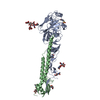
 Movie
Movie Controller


+ Open data

- Basic information
| Entry | Database: PDB / ID: 1boy | ||||||
|---|---|---|---|---|---|---|---|
| Title | EXTRACELLULAR REGION OF HUMAN TISSUE FACTOR | ||||||
 Components Components | HUMAN TISSUE FACTOR | ||||||
 Keywords Keywords | CLASS 2 CYTOKINE RECEPTOR / INITIATOR OF BLOOD COAGULATION IN VERTEBRATES / COFACTOR FOR FACTOR VIIA / GLYCOPROTEIN / BLOOD COAGULATION | ||||||
| Function / homology |  Function and homology information Function and homology informationactivation of plasma proteins involved in acute inflammatory response / activation of blood coagulation via clotting cascade / serine-type peptidase complex / positive regulation of platelet-derived growth factor receptor signaling pathway / NGF-stimulated transcription / cytokine receptor activity / positive regulation of positive chemotaxis / Extrinsic Pathway of Fibrin Clot Formation / positive regulation of TOR signaling / positive regulation of endothelial cell proliferation ...activation of plasma proteins involved in acute inflammatory response / activation of blood coagulation via clotting cascade / serine-type peptidase complex / positive regulation of platelet-derived growth factor receptor signaling pathway / NGF-stimulated transcription / cytokine receptor activity / positive regulation of positive chemotaxis / Extrinsic Pathway of Fibrin Clot Formation / positive regulation of TOR signaling / positive regulation of endothelial cell proliferation / positive regulation of interleukin-8 production / phospholipid binding / protein processing / cytokine-mediated signaling pathway / activation of cysteine-type endopeptidase activity involved in apoptotic process / positive regulation of angiogenesis / blood coagulation / collagen-containing extracellular matrix / protease binding / positive regulation of cell migration / external side of plasma membrane / positive regulation of gene expression / cell surface / extracellular space / membrane / plasma membraneSimilarity search - Function | ||||||
| Biological species |  Homo sapiens (human) Homo sapiens (human) | ||||||
| Method | X-RAY DIFFRACTION / SYNCHROTRON / Resolution: 2.2 Å | ||||||
 Authors Authors | Boys, C.W.G. / Tuddenham, E.G.D. / Harlos, K. | ||||||
 Citation Citation | Journal: Nature / Year: 1994 Title: Crystal structure of the extracellular region of human tissue factor. Authors: Harlos, K. / Martin, D.M. / O'Brien, D.P. / Jones, E.Y. / Stuart, D.I. / Polikarpov, I. / Miller, A. / Tuddenham, E.G. / Boys, C.W. #1: Journal: J.Mol.Biol. / Year: 1993Title: Crystallization and Preliminary X-Ray Analysis of Human Tissue Factor Extracellular Domain Authors: Boys, C.W. / Miller, A. / Harlos, K. / Martin, D.M. / Tuddenham, E.G. / O'Brien, D.P. | ||||||
| History |
|
- Structure visualization
| Structure viewer | Molecule: MolmilJmol/JSmol |
|---|
- Downloads & links
-Download
| PDBx/mmCIF format | 1boy.cif.gz | 53.4 KB | Display | PDBx/mmCIF format |
|---|---|---|---|---|
| PDB format | pdb1boy.ent.gz | 42 KB | Display | PDB format |
| PDBx/mmJSON format | 1boy.json.gz | Tree view | PDBx/mmJSON format | |
| Others |  Other downloads Other downloads |
-Validation report
| Arichive directory | https://data.pdbj.org/pub/pdb/validation_reports/bo/1boyftp://data.pdbj.org/pub/pdb/validation_reports/bo/1boy | HTTPS FTP |
|---|
-Related structure data
| Similar structure data |
|---|


-Links
 PDBj
PDBj


- Assembly
| Deposited unit | 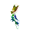
| ||||||||
|---|---|---|---|---|---|---|---|---|---|
| 1 | 
| ||||||||
| Unit cell |
|
-Components
| #1: Protein | / TF Mass: 24826.512 Da / Num. of mol.: 1 / Fragment: EXTRACELLULAR DOMAIN, RESIDUES 1 - 219 Source method: isolated from a genetically manipulated source Details: TYPE B CRYSTALS / Source: (gene. exp.) Homo sapiens (human) / Strain: W3110Description: PURIFIED BY MONOCLONAL ANTIBODY AFFINITY, REVERSED PHASE HPLC AND ISO-ELECTRIC FOCUSSING Gene: HUMAN TISSUE FACTOR CDNA CODON / Organ: BLOOD / Variant: TONA / Plasmid: PHGH4L (GENE 55:189-196, 1987)Gene (production host): HUMAN TISSUE FACTOR CDNA CODONS 1-119 Production host: PERIPLASMIC SECRETION (U.S PAT 4963,495) / References: UniProt: P13726 |
|---|---|
| #2: Water | ChemComp-HOH / Water Mass: 18.015 Da / Num. of mol.: 110 / Source method: isolated from a natural source / Formula: H2O Mass: 18.015 Da / Num. of mol.: 110 / Source method: isolated from a natural source / Formula: H2O |
-Experimental details
-Experiment
| Experiment | Method: X-RAY DIFFRACTION |
|---|
- Sample preparation
| Crystal | Density Matthews: 2.39 Å3/Da / Density % sol: 48 % Description: THE REDUNDANCY GIVEN ABOVE IS TO 2.2 ANGSTROMS RESOLUTION. | ||||||||||||||||||||
|---|---|---|---|---|---|---|---|---|---|---|---|---|---|---|---|---|---|---|---|---|---|
| Crystal grow | *PLUS pH: 7.5 / Method: vapor diffusion, hanging drop | ||||||||||||||||||||
| Components of the solutions | *PLUS
|
-Data collection
| Diffraction source | Source: SYNCHROTRON / Site: Photon Factory  / Beamline: BL-6A / Wavelength: 0.97 / Beamline: BL-6A / Wavelength: 0.97 |
|---|---|
| Detector | Type: FUJI / Detector: IMAGE PLATE / Date: Mar 1, 1994 |
| Radiation | Monochromatic (M) / Laue (L): M / Scattering type: x-ray |
| Radiation wavelength | Wavelength: 0.97 Å / Relative weight: 1 |
| Reflection | Num. obs: 12666 / % possible obs: 96 % / Observed criterion σ(I): 3 / Redundancy: 6.4 % / Rmerge(I) obs: 0.065 |
| Reflection | *PLUS Highest resolution: 2.2 Å |
- Processing
| Software |
| ||||||||||||||||||||||||||||||||||||||||||||||||||||||||||||
|---|---|---|---|---|---|---|---|---|---|---|---|---|---|---|---|---|---|---|---|---|---|---|---|---|---|---|---|---|---|---|---|---|---|---|---|---|---|---|---|---|---|---|---|---|---|---|---|---|---|---|---|---|---|---|---|---|---|---|---|---|---|
| Refinement | Resolution: 2.2→15 Å /
| ||||||||||||||||||||||||||||||||||||||||||||||||||||||||||||
| Refinement step | Cycle: LAST / Resolution: 2.2→15 Å
| ||||||||||||||||||||||||||||||||||||||||||||||||||||||||||||
| Refine LS restraints |
| ||||||||||||||||||||||||||||||||||||||||||||||||||||||||||||
| Software | *PLUS Name: X-PLOR / Classification: refinement | ||||||||||||||||||||||||||||||||||||||||||||||||||||||||||||
| Refinement | *PLUS | ||||||||||||||||||||||||||||||||||||||||||||||||||||||||||||
| Solvent computation | *PLUS | ||||||||||||||||||||||||||||||||||||||||||||||||||||||||||||
| Displacement parameters | *PLUS Biso mean: 48.9 Å2 |
