 Movie
Movie Controller

[English] 日本語
 Yorodumi
- PDB-1bol: THE CRYSTAL STRUCTURE OF RIBONUCLEASE RH FROM RHIZOPUS NIVEUS AT ... -
+ Open data

- Basic information
| Entry | Database: PDB / ID: 1bol | ||||||
|---|---|---|---|---|---|---|---|
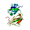
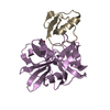
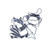
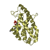
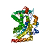
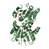
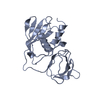
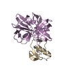
| Title | THE CRYSTAL STRUCTURE OF RIBONUCLEASE RH FROM RHIZOPUS NIVEUS AT 2.0 A RESOLUTION | ||||||
 Components Components | PROTEIN (RIBONUCLEASE RH) | ||||||
 Keywords Keywords |  HYDROLASE / RIBONUCLEASES HYDROLASE / RIBONUCLEASES | ||||||
| Function / homology |  Function and homology informationribonuclease T2 / ribonuclease T2 activity / lyase activity / RNA binding Function and homology informationribonuclease T2 / ribonuclease T2 activity / lyase activity / RNA bindingSimilarity search - Function | ||||||
| Biological species |  Rhizopus niveus (fungus) Rhizopus niveus (fungus) | ||||||
| Method | X-RAY DIFFRACTION / SYNCHROTRON / MIR / Resolution: 2 Å | ||||||
 Authors Authors | Kurihara, H. / Nakamura, K.T. | ||||||
 Citation Citation | Journal: J.Mol.Biol. / Year: 1996 Title: The crystal structure of ribonuclease Rh from Rhizopus niveus at 2.0 A resolution. Authors: Kurihara, H. / Nonaka, T. / Mitsui, Y. / Ohgi, K. / Irie, M. / Nakamura, K.T. | ||||||
| History |
|
- Structure visualization
| Structure viewer | Molecule: MolmilJmol/JSmol |
|---|
- Downloads & links
-Download
| PDBx/mmCIF format | 1bol.cif.gz | 48.2 KB | Display | PDBx/mmCIF format |
|---|---|---|---|---|
| PDB format | pdb1bol.ent.gz | 38.3 KB | Display | PDB format |
| PDBx/mmJSON format | 1bol.json.gz | Tree view | PDBx/mmJSON format | |
| Others |  Other downloads Other downloads |
-Validation report
| Arichive directory | https://data.pdbj.org/pub/pdb/validation_reports/bo/1bolftp://data.pdbj.org/pub/pdb/validation_reports/bo/1bol | HTTPS FTP |
|---|
-Related structure data
| Similar structure data |
|---|

-Links
 PDBj
PDBj- Assembly
| Deposited unit | 
| ||||||||
|---|---|---|---|---|---|---|---|---|---|
| 1 |
| ||||||||
| Unit cell |
|
-Components
| #1: Protein | Mass: 24096.203 Da / Num. of mol.: 1 / Source method: isolated from a natural source / Source: (natural) Rhizopus niveus (fungus) / References: UniProt: P08056, EC: 3.1.27.1 |
|---|
-Experimental details
-Experiment
| Experiment | Method: X-RAY DIFFRACTION / Number of used crystals: 2 |
|---|
- Sample preparation
| Crystal | Density Matthews: 2.6 Å3/Da / Density % sol: 45.46 % | |||||||||||||||||||||||||
|---|---|---|---|---|---|---|---|---|---|---|---|---|---|---|---|---|---|---|---|---|---|---|---|---|---|---|
| Crystal grow | pH: 6.7 / Details: pH 6.7 | |||||||||||||||||||||||||
| Crystal grow | *PLUS Temperature: 20 ℃ / Method: vapor diffusion, hanging drop | |||||||||||||||||||||||||
| Components of the solutions | *PLUS
|
-Data collection
| Diffraction | Mean temperature: 293 K |
|---|---|
| Diffraction source | Source: SYNCHROTRON / Site: Photon Factory  / Beamline: BL-6A / Wavelength: 1.5418 / Wavelength: 1.5418 Å / Beamline: BL-6A / Wavelength: 1.5418 / Wavelength: 1.5418 Å |
| Detector | Type: RIGAKU / Detector: IMAGE PLATE |
| Radiation | Monochromator: NI FILTER / Protocol: SINGLE WAVELENGTH / Monochromatic (M) / Laue (L): M / Scattering type: x-ray |
| Radiation wavelength | Wavelength: 1.5418 Å / Relative weight: 1 |
| Reflection | Resolution: 2→8 Å / Num. obs: 11745 / % possible obs: 78.2 % / Observed criterion σ(I): 2 / Redundancy: 2.5 % / Rmerge(I) obs: 0.0334 |
- Processing
| Software | Name: X-PLOR / Classification: refinement | ||||||||||||||||||||||||||||||||||||||||||||||||||||||||||||
|---|---|---|---|---|---|---|---|---|---|---|---|---|---|---|---|---|---|---|---|---|---|---|---|---|---|---|---|---|---|---|---|---|---|---|---|---|---|---|---|---|---|---|---|---|---|---|---|---|---|---|---|---|---|---|---|---|---|---|---|---|---|
| Refinement | Method to determine structure: MIR / Resolution: 2→8 Å / σ(F): 2
| ||||||||||||||||||||||||||||||||||||||||||||||||||||||||||||
| Refinement step | Cycle: LAST / Resolution: 2→8 Å
| ||||||||||||||||||||||||||||||||||||||||||||||||||||||||||||
| Refine LS restraints |
| ||||||||||||||||||||||||||||||||||||||||||||||||||||||||||||
| Refine LS restraints | *PLUS
|
