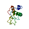
 Movie
Movie Controller


+ Open data

- Basic information
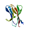
| Entry | Database: PDB / ID: 1blu | ||||||
|---|---|---|---|---|---|---|---|
| Title | STRUCTURE OF THE 2[4FE-4S] FERREDOXIN FROM CHROMATIUM VINOSUM | ||||||
 Components Components | FERREDOXIN | ||||||
 Keywords Keywords | ELECTRON TRANSPORT / FERREDOXIN | ||||||
| Function / homology |  Function and homology information Function and homology information | ||||||
| Biological species |  Allochromatium vinosum (bacteria) Allochromatium vinosum (bacteria) | ||||||
| Method | X-RAY DIFFRACTION / SYNCHROTRON / molecular replacement / Resolution: 2.1 Å | ||||||
 Authors Authors | Dauter, Z. / Wilson, K.S. / Sieker, L.C. / Moulis, J.M. | ||||||
 Citation Citation | Journal: Protein Sci. / Year: 1996 Title: Crystal structure of the 2[4Fe-4S] ferredoxin from Chromatium vinosum: evolutionary and mechanistic inferences for [3/4Fe-4S] ferredoxins. Authors: Moulis, J.M. / Sieker, L.C. / Wilson, K.S. / Dauter, Z. | ||||||
| History |
|
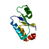
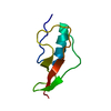
- Structure visualization
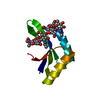
| Structure viewer | Molecule: MolmilJmol/JSmol |
|---|
- Downloads & links
-Download
| PDBx/mmCIF format | 1blu.cif.gz | 26.7 KB | Display | PDBx/mmCIF format |
|---|---|---|---|---|
| PDB format | pdb1blu.ent.gz | 19.5 KB | Display | PDB format |
| PDBx/mmJSON format | 1blu.json.gz | Tree view | PDBx/mmJSON format | |
| Others |  Other downloads Other downloads |
-Validation report
| Arichive directory | https://data.pdbj.org/pub/pdb/validation_reports/bl/1bluftp://data.pdbj.org/pub/pdb/validation_reports/bl/1blu | HTTPS FTP |
|---|
-Related structure data
| Related structure data |  1fdx S: Starting model for refinement |
|---|---|
| Similar structure data |


-Links
 PDBj
PDBj


- Assembly
| Deposited unit | 
| ||||||||
|---|---|---|---|---|---|---|---|---|---|
| 1 |
| ||||||||
| Unit cell |
|
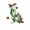
-Components
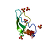
| #1: Protein | Mass: 9064.004 Da / Num. of mol.: 1 / Source method: isolated from a natural source / Source: (natural) Allochromatium vinosum (bacteria) / References: UniProt: P00208 | ||
|---|---|---|---|
| #2: Chemical | Iron–sulfur cluster  Mass: 351.640 Da / Num. of mol.: 2 / Source method: obtained synthetically / Formula: Fe4S4 Mass: 351.640 Da / Num. of mol.: 2 / Source method: obtained synthetically / Formula: Fe4S4#3: Water | ChemComp-HOH / | Water Mass: 18.015 Da / Num. of mol.: 85 / Source method: isolated from a natural source / Formula: H2O Mass: 18.015 Da / Num. of mol.: 85 / Source method: isolated from a natural source / Formula: H2O |
-Experimental details
-Experiment
| Experiment | Method: X-RAY DIFFRACTION |
|---|
- Sample preparation
| Crystal | Density Matthews: 3.32 Å3/Da / Density % sol: 63 % | ||||||||||||||||||||||||
|---|---|---|---|---|---|---|---|---|---|---|---|---|---|---|---|---|---|---|---|---|---|---|---|---|---|
| Crystal grow | pH: 6.5 Details: CRYSTALS WERE GROWN AT ROOM TEMPERATURE IN 0.1M TRIS-MALEATE BUFFER, PH 6.5 CONTAINING 2.9 M AMMONIUM SULPHATE AND 0.1 M NACL Temp details: room temp | ||||||||||||||||||||||||
| Crystal | *PLUS | ||||||||||||||||||||||||
| Crystal grow | *PLUS Temperature: 8 ℃ / Method: vapor diffusion, sitting drop | ||||||||||||||||||||||||
| Components of the solutions | *PLUS
|
-Data collection
| Diffraction source | Source: SYNCHROTRON / Site: EMBL/DESY, HAMBURG  / Beamline: X11 / Wavelength: 0.92 / Beamline: X11 / Wavelength: 0.92 |
|---|---|
| Detector | Type: MARRESEARCH / Detector: IMAGE PLATE / Date: Oct 1, 1993 |
| Radiation | Monochromatic (M) / Laue (L): M / Scattering type: x-ray |
| Radiation wavelength | Wavelength: 0.92 Å / Relative weight: 1 |
| Reflection | Resolution: 2.1→20 Å / Num. obs: 7178 / % possible obs: 97 % / Observed criterion σ(I): 0 / Redundancy: 5 % / Rmerge(I) obs: 0.081 |
- Processing
| Software |
| ||||||||||||||||||||||||||||||||||||||||||||||||||||||||||||||||||||||||||||||||||||
|---|---|---|---|---|---|---|---|---|---|---|---|---|---|---|---|---|---|---|---|---|---|---|---|---|---|---|---|---|---|---|---|---|---|---|---|---|---|---|---|---|---|---|---|---|---|---|---|---|---|---|---|---|---|---|---|---|---|---|---|---|---|---|---|---|---|---|---|---|---|---|---|---|---|---|---|---|---|---|---|---|---|---|---|---|---|
| Refinement | Method to determine structure: molecular replacement Starting model: 1FDX 1fdx Resolution: 2.1→20 Å / σ(F): 0 /
| ||||||||||||||||||||||||||||||||||||||||||||||||||||||||||||||||||||||||||||||||||||
| Refine analyze | Luzzati sigma a obs: 0.25 Å | ||||||||||||||||||||||||||||||||||||||||||||||||||||||||||||||||||||||||||||||||||||
| Refinement step | Cycle: LAST / Resolution: 2.1→20 Å
| ||||||||||||||||||||||||||||||||||||||||||||||||||||||||||||||||||||||||||||||||||||
| Refine LS restraints |
| ||||||||||||||||||||||||||||||||||||||||||||||||||||||||||||||||||||||||||||||||||||
| Software | *PLUS Name: PROLSQ / Classification: refinement | ||||||||||||||||||||||||||||||||||||||||||||||||||||||||||||||||||||||||||||||||||||
| Refinement | *PLUS Rfactor obs: 0.192 / Rfactor Rwork: 0.192 | ||||||||||||||||||||||||||||||||||||||||||||||||||||||||||||||||||||||||||||||||||||
| Solvent computation | *PLUS | ||||||||||||||||||||||||||||||||||||||||||||||||||||||||||||||||||||||||||||||||||||
| Displacement parameters | *PLUS |
