 Movie
Movie Controller

[English] 日本語
 Yorodumi
- PDB-1bjo: THE STRUCTURE OF PHOSPHOSERINE AMINOTRANSFERASE FROM E. COLI IN C... -
+ Open data

- Basic information
| Entry | Database: PDB / ID: 1bjo | ||||||
|---|---|---|---|---|---|---|---|
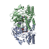
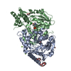
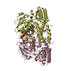
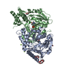
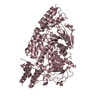
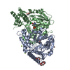
| Title | THE STRUCTURE OF PHOSPHOSERINE AMINOTRANSFERASE FROM E. COLI IN COMPLEX WITH ALPHA-METHYL-L-GLUTAMATE | ||||||
 Components Components | (PHOSPHOSERINE AMINOTRANSFERASE Phosphoserine transaminase) x 2 Phosphoserine transaminase) x 2 | ||||||
 Keywords Keywords | AMINOTRANSFERASE / L-SERINE BIOSYNTHESIS | ||||||
| Function / homology |  Function and homology information Function and homology informationlysine biosynthetic process via diaminopimelate and N-succinyl-2-amino-6-ketopimelate / phosphoserine transaminase / O-phospho-L-serine:2-oxoglutarate aminotransferase activity / pyridoxal phosphate biosynthetic process / pyridoxine biosynthetic process / L-serine metabolic process / L-serine biosynthetic process / pyridoxal phosphate binding / protein homodimerization activity / cytosol / cytoplasmSimilarity search - Function | ||||||
| Biological species |  Escherichia coli (E. coli) Escherichia coli (E. coli) | ||||||
| Method | X-RAY DIFFRACTION / DIFFERENCE FOURIER / Resolution: 2.8 Å | ||||||
 Authors Authors | Hester, G. / Stark, W. / Jansonius, J.N. | ||||||
 Citation Citation | Journal: J.Mol.Biol. / Year: 1999 Title: Crystal structure of phosphoserine aminotransferase from Escherichia coli at 2.3 A resolution: comparison of the unligated enzyme and a complex with alpha-methyl-l-glutamate. Authors: Hester, G. / Stark, W. / Moser, M. / Kallen, J. / Markovic-Housley, Z. / Jansonius, J.N. #1: Journal: Enzymes Dependent on Pyridoxal Phosphate and Other Carbonyl Compounds as Cofactors: Proceedings of the 8Th International Symposium on Vitamin B6 and Carbonyl CatalysisYear: 1991 Title: The Three Dimensional Structure of Phosphoserine Aminotransferase from Escherichia Coli Authors: Stark, W. / Kallen, J. / Markovic-Housley, Z. / Fol, B. / Kania, M. / Jansonius, J.N. #2: Journal: Biochemistry of Vitamin B6: Proceedings of the 7Th International Congress on Chemical and Biological Aspects of Vitamin B6 Catalysis (in: Iub Symp. Ser., V.166)Year: 1987 Title: Crystallographic and Solution Studies on Phosphoserine Aminotransferase (Psat) from E. Coli Authors: Kallen, J. / Kania, M. / Markovic-Housley, Z. / Vincent, M.G. / Jansonius, J.N. | ||||||
| History |
|
- Structure visualization
| Structure viewer | Molecule: MolmilJmol/JSmol |
|---|
- Downloads & links
-Download
| PDBx/mmCIF format | 1bjo.cif.gz | 135 KB | Display | PDBx/mmCIF format |
|---|---|---|---|---|
| PDB format | pdb1bjo.ent.gz | 111.9 KB | Display | PDB format |
| PDBx/mmJSON format | 1bjo.json.gz | Tree view | PDBx/mmJSON format | |
| Others |  Other downloads Other downloads |
-Validation report
| Arichive directory | https://data.pdbj.org/pub/pdb/validation_reports/bj/1bjoftp://data.pdbj.org/pub/pdb/validation_reports/bj/1bjo | HTTPS FTP |
|---|
-Related structure data


-Links
 PDBj
PDBj- Assembly
| Deposited unit | 
| ||||||||
|---|---|---|---|---|---|---|---|---|---|
| 1 |
| ||||||||
| Unit cell |
| ||||||||
| Noncrystallographic symmetry (NCS) | NCS oper: (Code: given Matrix: (0.45212, -0.73722, 0.50209), Vector : |
-Components
| #1: Protein | Phosphoserine transaminase / PSAT Mass: 39709.047 Da / Num. of mol.: 1 Source method: isolated from a genetically manipulated source Details: IN CHAIN A AN EXTERNAL ALDIMINE INTERMEDIATE BETWEEN PYRIDOXAL-5'-PHOSPHATE AND ALPHA-METHYL-L-GLUTAMATE IS NON-COVALENTLY BOUND WHILE IN CHAIN B A PYRIDOXAL-5'-PHOSPHATE MOLECULE IS ...Details: IN CHAIN A AN EXTERNAL ALDIMINE INTERMEDIATE BETWEEN PYRIDOXAL-5'-PHOSPHATE AND ALPHA-METHYL-L-GLUTAMATE IS NON-COVALENTLY BOUND WHILE IN CHAIN B A PYRIDOXAL-5'-PHOSPHATE MOLECULE IS COVALENTLY BOUND IN AN ALDIMINE LINKAGE TO A LYSINE SIDE CHAIN Source: (gene. exp.) Escherichia coli (E. coli) / Gene: SERCReferences: PIR: S28806, UniProt: P23721*PLUS, phosphoserine transaminase |
|---|---|
| #2: Protein | Phosphoserine transaminase / PSAT Mass: 39937.164 Da / Num. of mol.: 1 Source method: isolated from a genetically manipulated source Details: IN CHAIN A AN EXTERNAL ALDIMINE INTERMEDIATE BETWEEN PYRIDOXAL-5'-PHOSPHATE AND ALPHA-METHYL-L-GLUTAMATE IS NON-COVALENTLY BOUND WHILE IN CHAIN B A PYRIDOXAL-5'-PHOSPHATE MOLECULE IS ...Details: IN CHAIN A AN EXTERNAL ALDIMINE INTERMEDIATE BETWEEN PYRIDOXAL-5'-PHOSPHATE AND ALPHA-METHYL-L-GLUTAMATE IS NON-COVALENTLY BOUND WHILE IN CHAIN B A PYRIDOXAL-5'-PHOSPHATE MOLECULE IS COVALENTLY BOUND IN AN ALDIMINE LINKAGE TO A LYSINE SIDE CHAIN Source: (gene. exp.) Escherichia coli (E. coli) / Gene: SERCReferences: PIR: S28806, UniProt: P23721*PLUS, phosphoserine transaminase |
| #3: Chemical | ChemComp-PLP / Pyridoxal phosphate  Mass: 247.142 Da / Num. of mol.: 1 / Source method: obtained synthetically / Formula: C8H10NO6P Mass: 247.142 Da / Num. of mol.: 1 / Source method: obtained synthetically / Formula: C8H10NO6P |
| #4: Chemical | ChemComp-GAM /   Mass: 161.156 Da / Num. of mol.: 1 / Source method: obtained synthetically / Formula: C6H11NO4 Mass: 161.156 Da / Num. of mol.: 1 / Source method: obtained synthetically / Formula: C6H11NO4 |
| #5: Water | ChemComp-HOH / Water Mass: 18.015 Da / Num. of mol.: 50 / Source method: isolated from a natural source / Formula: H2O Mass: 18.015 Da / Num. of mol.: 50 / Source method: isolated from a natural source / Formula: H2O |
-Experimental details
-Experiment
| Experiment | Method: X-RAY DIFFRACTION / Number of used crystals: 1 |
|---|
- Sample preparation
| Crystal | Density Matthews: 2.7 Å3/Da / Density % sol: 52 % | ||||||||||||
|---|---|---|---|---|---|---|---|---|---|---|---|---|---|
| Crystal grow | Temperature: 277 K / Method: vapor diffusion, hanging drop Details: PSAT HAS BEEN CRYSTALLIZED AT 4-7 DEGREES CELSIUS BY THE HANGING DROP METHOD, USING PEG4000 AS A PRECIPITANT, BUFFERED WITH SODIUM ACETATE TO PH 7.2 IN THE DROP AND PH 5.6 IN THE RESERVOIR. ...Details: PSAT HAS BEEN CRYSTALLIZED AT 4-7 DEGREES CELSIUS BY THE HANGING DROP METHOD, USING PEG4000 AS A PRECIPITANT, BUFFERED WITH SODIUM ACETATE TO PH 7.2 IN THE DROP AND PH 5.6 IN THE RESERVOIR. THE PH-GRADIENT WAS ESSENTIAL FOR THE CRYSTALLIZATION., vapor diffusion - hanging drop, temperature 277K PH range: 5.6-7.2 | ||||||||||||
| Crystal grow | *PLUS Temperature: 4-7 ℃ / Method: vapor diffusion, hanging drop / Details: pH7.2 in the drop and pH5.6 in the reservoir | ||||||||||||
| Components of the solutions | *PLUS
|
-Data collection
| Diffraction | Mean temperature: 277 K |
|---|---|
| Diffraction source | Source: ROTATING ANODE / Type: ELLIOTT GX-20 / Wavelength: 1.5418 |
| Detector | Type: ENRAF-NONIUS FAST / Detector: DIFFRACTOMETER / Date: Jul 1, 1991 |
| Radiation | Monochromatic (M) / Laue (L): M / Scattering type: x-ray |
| Radiation wavelength | Wavelength: 1.5418 Å / Relative weight: 1 |
| Reflection | Resolution: 2.8→39.8 Å / Num. obs: 19908 / % possible obs: 91.8 % / Observed criterion σ(I): 0 / Redundancy: 2.7 % / Biso Wilson estimate: 45.4 Å2 / Rsym value: 0.094 / Net I/σ(I): 9.8 |
| Reflection shell | Resolution: 2.8→2.95 Å / Redundancy: 1.8 % / Mean I/σ(I) obs: 2.8 / Rsym value: 0.402 / % possible all: 68.4 |
| Reflection | *PLUS Rmerge(I) obs: 0.094 / Biso Wilson estimate: 29.1 Å2 |
| Reflection shell | *PLUS % possible obs: 68.4 % / Rmerge(I) obs: 0.402 |
- Processing
| Software |
| ||||||||||||||||||||||||||||||||||||||||||||||||||||||||||||
|---|---|---|---|---|---|---|---|---|---|---|---|---|---|---|---|---|---|---|---|---|---|---|---|---|---|---|---|---|---|---|---|---|---|---|---|---|---|---|---|---|---|---|---|---|---|---|---|---|---|---|---|---|---|---|---|---|---|---|---|---|---|
| Refinement | Method to determine structure: DIFFERENCE FOURIER / Resolution: 2.8→39.8 Å / Rfactor Rfree error: 0.008 / Data cutoff high absF: 10000000 / Data cutoff low absF: 0.001 / Isotropic thermal model: GROUP / Cross valid method: THROUGHOUT / σ(F): 0 / Details: BULK SOLVENT MODEL USED
| ||||||||||||||||||||||||||||||||||||||||||||||||||||||||||||
| Displacement parameters | Biso mean: 33.5 Å2
| ||||||||||||||||||||||||||||||||||||||||||||||||||||||||||||
| Refine analyze |
| ||||||||||||||||||||||||||||||||||||||||||||||||||||||||||||
| Refinement step | Cycle: LAST / Resolution: 2.8→39.8 Å
| ||||||||||||||||||||||||||||||||||||||||||||||||||||||||||||
| Refine LS restraints |
| ||||||||||||||||||||||||||||||||||||||||||||||||||||||||||||
| Refine LS restraints NCS | Rms dev Biso : 1.5 Å2 / Rms dev position: 0.48 Å / Weight position: 500 | ||||||||||||||||||||||||||||||||||||||||||||||||||||||||||||
| LS refinement shell | Resolution: 2.8→2.9 Å / Rfactor Rfree error: 0.05 / Total num. of bins used: 10
| ||||||||||||||||||||||||||||||||||||||||||||||||||||||||||||
| Xplor file |
| ||||||||||||||||||||||||||||||||||||||||||||||||||||||||||||
| Software | *PLUS Name: X-PLOR / Version: 3.851 / Classification: refinement | ||||||||||||||||||||||||||||||||||||||||||||||||||||||||||||
| Refinement | *PLUS | ||||||||||||||||||||||||||||||||||||||||||||||||||||||||||||
| Solvent computation | *PLUS | ||||||||||||||||||||||||||||||||||||||||||||||||||||||||||||
| Displacement parameters | *PLUS | ||||||||||||||||||||||||||||||||||||||||||||||||||||||||||||
| Refine LS restraints | *PLUS
| ||||||||||||||||||||||||||||||||||||||||||||||||||||||||||||
| LS refinement shell | *PLUS Rfactor obs: 0.315 |
