 Movie
Movie Controller


+ Open data

- Basic information
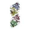
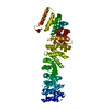
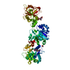
| Entry | Database: PDB / ID: 1bg9 | |||||||||
|---|---|---|---|---|---|---|---|---|---|---|
| Title | BARLEY ALPHA-AMYLASE WITH SUBSTRATE ANALOGUE ACARBOSE | |||||||||
 Components Components | 1,4-ALPHA-D-GLUCAN GLUCANOHYDROLASE | |||||||||
 Keywords Keywords |  HYDROLASE / O-GLYCOSYL HYDROLASE / O-GLYCOSYL | |||||||||
| Function / homology |  Function and homology information Function and homology informationstarch catabolic process / alpha-amylase / alpha-amylase activity / calcium ion bindingSimilarity search - Function | |||||||||
| Biological species |  Hordeum vulgare (barley) Hordeum vulgare (barley) | |||||||||
| Method | X-RAY DIFFRACTION / REFINEMENT DIFFERENCE FOURIER / Resolution: 2.8 Å | |||||||||
 Authors Authors | Kadziola, A. / Haser, R. | |||||||||
 Citation Citation | Journal: J.Mol.Biol. / Year: 1998 Title: Molecular structure of a barley alpha-amylase-inhibitor complex: implications for starch binding and catalysis. Authors: Kadziola, A. / Sogaard, M. / Svensson, B. / Haser, R. #1: Journal: J.Mol.Biol. / Year: 1994Title: Crystal and Molecular Structure of Barley Alpha-Amylase Authors: Kadziola, A. / Abe, J.I. / Svensson, B. / Haser, R. | |||||||||
| History |
|
- Structure visualization
| Structure viewer | Molecule: MolmilJmol/JSmol |
|---|
- Downloads & links
-Download
| PDBx/mmCIF format | 1bg9.cif.gz | 94.7 KB | Display | PDBx/mmCIF format |
|---|---|---|---|---|
| PDB format | pdb1bg9.ent.gz | 74.1 KB | Display | PDB format |
| PDBx/mmJSON format | 1bg9.json.gz | Tree view | PDBx/mmJSON format | |
| Others |  Other downloads Other downloads |
-Validation report
| Arichive directory | https://data.pdbj.org/pub/pdb/validation_reports/bg/1bg9ftp://data.pdbj.org/pub/pdb/validation_reports/bg/1bg9 | HTTPS FTP |
|---|
-Related structure data
| Related structure data |  1amyS S: Starting model for refinement |
|---|---|
| Similar structure data |


-Links
 PDBj
PDBj

- Assembly
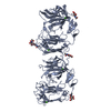
| Deposited unit | 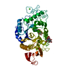
| ||||||||
|---|---|---|---|---|---|---|---|---|---|
| 1 | 
| ||||||||
| Unit cell |
|
-Components
| #1: Protein | Mass: 44986.438 Da / Num. of mol.: 1 / Source method: isolated from a natural source / Source: (natural) Hordeum vulgare (barley) / Organ: GERMINATED SEEDS / Variant: CULTIVAR MENUET / References: UniProt: P04063, alpha-amylase | ||||
|---|---|---|---|---|---|
| #2: Polysaccharide | 4,6-dideoxy-4-{[(1S,5R,6S)-3-formyl-5,6-dihydroxy-4-oxocyclohex-2-en-1-yl]amino}-alpha-D-xylo-hex-5- ...4,6-dideoxy-4-{[(1S,5R,6S)-3-formyl-5,6-dihydroxy-4-oxocyclohex-2-en-1-yl]amino}-alpha-D-xylo-hex-5-enopyranose-(1-4)-beta-D-glucopyranose Type: oligosaccharide / Mass: 477.417 Da / Num. of mol.: 1Source method: isolated from a genetically manipulated source | ||||
| #3: Chemical |   Mass: 40.078 Da / Num. of mol.: 3 / Source method: obtained synthetically / Formula: Ca Mass: 40.078 Da / Num. of mol.: 3 / Source method: obtained synthetically / Formula: Ca#4: Sugar | ChemComp-AF1 / |   Type: D-saccharide, beta linking / Mass: 321.324 Da / Num. of mol.: 1 Type: D-saccharide, beta linking / Mass: 321.324 Da / Num. of mol.: 1Source method: isolated from a genetically manipulated source Formula: C13H23NO8 #5: Water | ChemComp-HOH / | Water Mass: 18.015 Da / Num. of mol.: 148 / Source method: isolated from a natural source / Formula: H2O Mass: 18.015 Da / Num. of mol.: 148 / Source method: isolated from a natural source / Formula: H2O |
-Experimental details
-Experiment
| Experiment | Method: X-RAY DIFFRACTION / Number of used crystals: 1 |
|---|
- Sample preparation
| Crystal | Density Matthews: 4.67 Å3/Da / Density % sol: 75 % Description: COMPLEX BETWEEN BARLEY ALPHA-AMYLASE AND SUBSTRATE ANALOGUE ACARBOSE | ||||||||||||||||||||||||||||||
|---|---|---|---|---|---|---|---|---|---|---|---|---|---|---|---|---|---|---|---|---|---|---|---|---|---|---|---|---|---|---|---|
| Crystal grow | pH: 6.7 / Details: pH 6.7 | ||||||||||||||||||||||||||||||
| Crystal | *PLUS Density % sol: 75 % | ||||||||||||||||||||||||||||||
| Crystal grow | *PLUS Method: vapor diffusion, hanging drop / Details: Svensson, B., (1987) J. Biol. Chem., 262, 13682. | ||||||||||||||||||||||||||||||
| Components of the solutions | *PLUS
|
-Data collection
| Diffraction | Mean temperature: 288 K |
|---|---|
| Diffraction source | Source: ROTATING ANODE / Type: RIGAKU RUH2R / Wavelength: 1.5418 |
| Detector | Type: SIEMENS / Detector: AREA DETECTOR / Date: Apr 1, 1992 |
| Radiation | Monochromator: GRAPHITE(002) / Monochromatic (M) / Laue (L): M / Scattering type: x-ray |
| Radiation wavelength | Wavelength: 1.5418 Å / Relative weight: 1 |
| Reflection | Highest resolution: 2.729 Å / Num. obs: 17688 / % possible obs: 90.5 % / Observed criterion σ(I): 1 / Redundancy: 2.9 % / Rmerge(I) obs: 0.092 / Rsym value: 0.092 / Net I/σ(I): 16.6 |
| Reflection shell | Resolution: 2.729→2.9 Å / Redundancy: 1.5 % / Rmerge(I) obs: 0.27 / Mean I/σ(I) obs: 1.7 / Rsym value: 0.27 / % possible all: 44.8 |
| Reflection | *PLUS Num. measured all: 61039 |
| Reflection shell | *PLUS % possible obs: 44.8 % |
- Processing
| Software |
| ||||||||||||||||||||||||||||||||||||||||||||||||||||||||||||||||||||||||||||||||
|---|---|---|---|---|---|---|---|---|---|---|---|---|---|---|---|---|---|---|---|---|---|---|---|---|---|---|---|---|---|---|---|---|---|---|---|---|---|---|---|---|---|---|---|---|---|---|---|---|---|---|---|---|---|---|---|---|---|---|---|---|---|---|---|---|---|---|---|---|---|---|---|---|---|---|---|---|---|---|---|---|---|
| Refinement | Method to determine structure: REFINEMENT DIFFERENCE FOURIER Starting model: 1AMY Resolution: 2.8→10 Å Cross valid method: THE VERSION OF X-PLOR (2.1) ORIGINALLY USED FOR REFINEMENT THE FREE R TEST WAS NOT AN OPTION. THEREFORE WE HAVE GIVEN THE OVERALL R FACTOR (TEST+WORKING) INSTEAD OF (WORK) ...Cross valid method: THE VERSION OF X-PLOR (2.1) ORIGINALLY USED FOR REFINEMENT THE FREE R TEST WAS NOT AN OPTION. THEREFORE WE HAVE GIVEN THE OVERALL R FACTOR (TEST+WORKING) INSTEAD OF (WORK) TOGETHER WITH AN ESTIMATE OF THE FREE R (TEST) VALUE BASED ON A SIMULATED ANNEALING REFINEMENT WITH A NEWER VERSION OF X-PLOR (3.1). σ(F): 1
| ||||||||||||||||||||||||||||||||||||||||||||||||||||||||||||||||||||||||||||||||
| Displacement parameters | Biso mean: 20.1 Å2 | ||||||||||||||||||||||||||||||||||||||||||||||||||||||||||||||||||||||||||||||||
| Refine analyze |
| ||||||||||||||||||||||||||||||||||||||||||||||||||||||||||||||||||||||||||||||||
| Refinement step | Cycle: LAST / Resolution: 2.8→10 Å
| ||||||||||||||||||||||||||||||||||||||||||||||||||||||||||||||||||||||||||||||||
| Refine LS restraints |
| ||||||||||||||||||||||||||||||||||||||||||||||||||||||||||||||||||||||||||||||||
| LS refinement shell | Resolution: 2.8→2.92 Å / Total num. of bins used: 8
| ||||||||||||||||||||||||||||||||||||||||||||||||||||||||||||||||||||||||||||||||
| Xplor file |
| ||||||||||||||||||||||||||||||||||||||||||||||||||||||||||||||||||||||||||||||||
| Software | *PLUS Name: X-PLOR / Version: 2.1,3.1 / Classification: refinement | ||||||||||||||||||||||||||||||||||||||||||||||||||||||||||||||||||||||||||||||||
| Refine LS restraints | *PLUS
|
